Astragalus Root (5:1 extract)

Astragalus Root (5:1 extract)

Astragalus is a perennial plant that is native to parts of China, Mongolia, and Korea. The root is harvested from four year old plants and used in combination with other herbs. It is considered an adaptogen, a substance that helps the body cope with a variety of stress factors. The root of astragalus contains an active compound called cycloastragenol (TAT2) which is known to possess anti-aging properties

Benefits of Astragalus Root

An overwhelming body of high quality research and ongoing studies support the many health benefits of astragalus root. Taking astragalus root supplements can give you the following proven health benefits:

Boosts Immune Function

A study out of Beijing found that astragalus root has the ability to control cells of the immune system such as T-helper cells 1 and 2, essentially regulating the body’s immune responses.[1] In another study, researchers found that astragalus can boost resistance to infections, thus, preventing the development of complications and other serious health conditions.[2]

Fights Cancer

Results from two high quality studies show that a substance known as astragaloside from astragalus root appears to have anti-cancer properties.[3-4] Researchers found that astragaloside inhibits the growth and reproduction of gastrointestinal cancer cells, liver cancer cells, and breast cancer cells, by affecting different signaling pathways involved in cancer cell cycle.

Protects the Heart

The flavonoids present in astragalus are highly potent antioxidants that help prevent the accumulation of harmful plaques in arteries and narrowing of the walls of blood vessels. According to a 2014 study published in the Chinese Journal of Integrative Medicine, injection of astragalus in addition to conventional treatment for heart inflammation makes treatment more effective.[5] 

Improves Blood Sugar Levels

A 2013 study published in the Journal of Evidenced-Based Complementary and Alternative Medicine found that the saponins, flavonoids and polysaccharides present in astragalus root were effective in treating and regulating type 1 and 2 diabetes.[6] Researchers found that these substances improve the body’s response to the effects of insulin, thus, maintaining blood sugar levels within normal limits.  

Slows the Aging Process

A 2012 study published in the International Journal of Molecular Sciences found that the

polysaccharides in astragalus root has anti-aging properties.[7] According to researchers, these polysaccharides seek reactive oxygen species (ROS) and neutralize their harmful effect on the body. Scientists believe that increased ROS levels accelerate the aging process.

Accelerates Wound Healing

In a 2012 study conducted by the researchers at the Institute of Pharmaceutics at Zhejiang University, it was concluded that wound treatment with astragaloside IV (the active ingredient in dried astragalus root) increased the recovery rate as much as 2 to 3 times in just 48 hours.[8] In addition, the treatment was associated with lesser scarring.

References:

  1. Huang LF, Yao YM, Li JF, et al. The effect of Astragaloside IV on immune function of regulatory T cell mediated by high mobility group box 1 protein in vitro. Fitoterapia. 2012;83(8):1514-22.
  2. Block KI, Mead MN. Immune system effects of echinacea, ginseng, and astragalus: a review. Integr Cancer Ther. 2003;2(3):247-67.
  3. Auyeung KK, Han QB, Ko JK. Astragalus membranaceus: A Review of its Protection Against Inflammation and Gastrointestinal Cancers. Am J Chin Med. 2016;44(1):1-22.
  4. Huang C, Xu D, Xia Q, Wang P, Rong C, Su Y. Reversal of P-glycoprotein-mediated multidrug resistance of human hepatic cancer cells by Astragaloside II. J Pharm Pharmacol. 2012;64(12):1741-50.
  5. Piao YL, Liang XC. Astragalus membranaceus injection combined with conventional treatment for viral myocarditis: a systematic review of randomized controlled trials. Chin J Integr Med. 2014;20(10):787-91.
  6. Agyemang K, Han L, Liu E, Zhang Y, Wang T, Gao X. Recent Advances in Astragalus membranaceus Anti-Diabetic Research: Pharmacological Effects of Its Phytochemical Constituents. Evidence-based Complementary and Alternative Medicine : eCAM. 2013;2013:654643. doi:10.1155/2013/654643.
  7. Li X-T, Zhang Y-K, Kuang H-X, et al. Mitochondrial Protection and Anti-aging Activity of Astragalus Polysaccharides and Their Potential Mechanism. International Journal of Molecular Sciences. 2012;13(2):1747-1761. doi:10.3390/ijms13021747.
  8. Chen X, Peng LH, Li N, et al. The healing and anti-scar effects of astragaloside IV on the wound repair in vitro and in vivo. J Ethnopharmacol. 2012;139(3):721-7.

Aspergillopepsin

Aspergillopepsin

Aspergillopepsin, also known as Aspergillus acid protease or Aspergillus acid proteinase, is a proteolytic enzyme. This enzyme aids in the proper digestion of proteins and carbohydrates so that they will be easily converted into glucose and be used effectively by every cell in the body. This enzyme is found in a variety of spore-forming fungi known as Aspergillus species. You can also get a decent amount of aspergillopepsin in the form of capsules and tablets in many health food stores.

Benefits of Aspergillopepsin

Taking aspergillopepsin has a lot of proven health benefits to offer. Among them are the following:

Protects Against Free Radical Damage

The antioxidant activities of Aspergillus species are extensively reported in several studies. It is postulated that their antioxidant properties come from their abilities to hunt free radicals and inactivate them to prevent further cellular damage.[1] This effect can help slow the signs of aging and prevent a wide array of diseases related to free radical damage.

Treats Celiac Disease

Celiac disease or sprue is an inheritable, life-long disease that is characterized by an inflammatory reaction to dietary gluten, a protein found in wheat, rye, barley, and other grains. The hallmark of the disease is intestinal damage resulting to malabsorption of nutrients, vitamins and minerals. If untreated, celiac disease can lead to various complications including anemia, neurological problems, cancer, bone diseases, and other inflammatory disorders. Interestingly, one clinical trial found that aspergillopepsin supplementation in patients with celiac disease provided relief from symptoms related to inadvertent gluten exposure.[2] This therapeutic effect is related to the ability of aspergillopepsin to digest gluten effectively.

Improves Energy Levels

Malabsorption of nutrients, vitamins and minerals caused by gluten intolerance can deplete one’s energy levels. Since a gluten-free diet can be deficient in fiber and other vitamins and minerals, scientists recommend oral aspergillopepsin supplements in patients with gluten intolerance to get the nutrients and energy they need.[3] In addition, since aspergillopepsin is a potent digestive enzyme, it aids in the proper digestion of starch and other carbohydrate-rich foods so that your body has sufficient energy stores.

Maintains a Healthy Digestive System

Taking aspergillopepsin can help improve digestive health by preventing constipation and normalizing bowel movements. Results from one study show that aspergillopepsin from Aspergillus oryzae stimulates growth of some bacteria that digest fiber.[4] This finding is highly significant since proper absorption of fiber can help improve bowel regularity. In addition, aspergillopepsin’s ability to stimulate the growth of beneficial bacteria can help restore the delicate balance of good and bad bacteria in the digestive tract.

Lowers High Cholesterol Levels

Aspergillopepsin’s benefits go beyond digestive health. Consuming this health-enhancing enzyme can help bring down high cholesterol levels. In fact, one of the most well-known Aspergillus products is lovastatin, the cholesterol-reducing small molecule which was originally isolated from Aspergillus terreus.[5]

References:

  1. Ruann Janser Soares de Castro and Helia Harumi Sato, “Protease from Aspergillus oryzae: Biochemical Characterization and Application as a Potential Biocatalyst for Production of Protein Hydrolysates with Antioxidant Activities,” Journal of Food Processing, vol. 2014, Article ID 372352, 11 pages, 2014. doi:10.1155/2014/372352.
  2. Ehren J, Morón B, Martin E, Bethune MT, Gray GM, Khosla C. A food-grade enzyme preparation with modest gluten detoxification properties. PLoS ONE. 2009;4(7):e6313.
  3. Savvateeva LV, Zamyatnin AA. Prospects of Developing Medicinal Therapeutic Strategies and Pharmaceutical Design for Effective Gluten Intolerance Treatment. Curr Pharm Des. 2016;22(16):2439-49.
  4. Beharka AA, Nagaraja TG. Effect of Aspergillus oryzae extract alone or in combination with antimicrobial compounds on ruminal bacteria. J Dairy Sci. 1998;81(6):1591-8.
  5. Available at:http://www.sciencedirect.com/science/article/pii/S0960982213000249. Accessed March 13, 2017.

Ashitaba

Ashitaba

Ashitaba is a tender herb grown in Japan. It’s a member of the Angelica family, and goes by the common name “leaves of tomorrow”.  The roots, stems, and leaves are used in traditional herbal medicine, mostly to treat acid reflux disease and stomach problems. Ashitaba contains an abundance of beta carotene, an important antioxidant, as well as fiber and potassium. It also contains vitamins K and E, as well as other B vitamins and trace minerals. Because of its high nutritional value, people take ashitaba supplements in order to improve their overall health.

Benefits of Ashitaba

A large body of high quality research supports the many health benefits of taking ashitaba supplements. Among them are the following:

Improves Body Composition

Taking ashitaba supplements can help you lose significant amount of weight and achieve a lean body. According to a Japanese study conducted in 2012, researchers found that ingestion of ashitaba green juice at a dose of 6.2 grams per day for 8 weeks resulted in significant reduction in body weight, body fat percentage, and body mass index (BMI) in 9 adult subjects with metabolic syndrome.[1]

Fights Cancer

Cancer patients undergoing chemotherapy normally experience hair loss in different body areas and weakening of the immune system, resulting in an increased susceptibility to a wide array of infections. This is because chemotherapeutic drugs destroy all rapidly dividing cells including normal cells of the hair follicles and immune system. Interestingly, results from two laboratory studies found that ashitaba has anti-tumor properties and it was able to suppress the growth of cancer cells by inducing programmed cell death.[2-3] Also, the results are fascinating because ashitaba was able to kill all malignant cells but it has no toxic effect on normal cells, suggesting that ashitaba supplementation in cancer patients may help reduce adverse side effects caused by chemotherapy.

Fights Depression

According to a 2013 study published in the Journal of Applied Pharmacology, ashitaba contains antidepressant substances such as prenylated chalcones, xanthoangelol and 4-hydroxyderricin.[4] These substances work by inhibiting monoamine oxidase (triggers depression) in the brain.

Protects the Liver

Previous studies reported that the extracts of ashitaba have potent antioxidant and anti-inflammatory properties that can help improve some markers of liver function in habitual alcohol drinkers.[5] Researchers found that heavy drinkers who took ashitaba supplements experienced significant improvement in liver function tests compared to those who took placebo.    

Fights Infection

Ashitaba shows evidence of strong antibacterial properties. According to a study conducted by the Pharmaceutical Society of Japan in 1999, xanthoangelol (I) and 4-hydroxyderricin (II) isolated from the root of ashitaba showed strong antibacterial activity against different species of disease-causing bacteria.[6] Similarly, the Kangweon National University in Korea reported that ashitaba along with other Umbelliferae plants were able to inhibit the growth of disease-causing bacteria.[7] In another study published in 2008 in the International Journal of Medicinal Mushrooms, researchers found that ashitaba extract was able to inhibit the growth of Methicillin-resistant Staphylococcus aureus (MRSA), a bacterium that is responsible for several difficult-to-treat infections in humans.[8]

References:

  1. Ohnogi H, Hayami S, Kudo Y, Enoki T. Efficacy and Safety of Ashitaba (Angelica keiskei) on the Patients and Candidates with Metabolic Syndrome: A Pilot Study. Jpn J. Complement. Alternat. Med. 2012;9(1):49–55.
  2. Okuyama T, Takata M, Takayasu J, et al. Anti-tumor-promotion by principles obtained from Angelica keiskei. Planta Med. 1991;57(3):242-6.
  3. Nishimura R, Tabata K, Arakawa M, et al. Isobavachalcone, a chalcone constituent of Angelica keiskei, induces apoptosis in neuroblastoma. Biol Pharm Bull. 2007;30(10):1878-83.
  4. Kim JH, Son YK, Kim GH, Hwang KH. Xanthoangelol and 4-Hydroxyderricin Are the Major Active Principles of the Inhibitory Activities against Monoamine Oxidases on Angelica keiskei K. Biomol Ther (Seoul). 2013;21(3):234-40.
  5. Noh HM, Ahn EM, Yun JM, Cho BL, Paek YJ. Angelica keiskei Koidzumi extracts improve some markers of liver function in habitual alcohol drinkers: a randomized double-blind clinical trial. J Med Food. 2015;18(2):166-72.
  6. Available at: http://ci.nii.ac.jp/naid/130003945577/. Accessed March 25, 2017.
  7. Kim CM, Heo MY, Kim HP, Sin KS, Pachaly P. Pharmacological activities of water extracts of Umbelliferae plants. Arch Pharm Res. 1991;14(1):87-92.
  8. Available at: http://www.dl.begellhouse.com/journals/708ae68d64b17c52,74677e1376d0a2ef,2c29695069ad0f3c.html. Accessed March 25, 2017.

Artichoke

Artichoke

Artichoke belongs to the sunflower family of vegetables and is actually considered a variety of thistle. Aside from being one of the favorite winter season edible flower buds used in international cuisines, artichoke also has medicinal properties because it is highly rich in fiber, vitamin C, vitamin B1, vitamin B2, vitamin B3, vitamin B6, vitamin B9, calcium, iron, zinc, manganese, sodium, potassium, and many other important nutrients!

Benefits of Artichoke

This nutrient powerhouse has diverse health benefits which are backed by high quality research. Among them are the following:

Weight Loss

Artichoke extract supplementation has been shown to reduce food intake in obese persons without highly abnormal parameters.[1] When subjects were given 200 mg of artichoke extract along with a low-caloric diet over 8 weeks, the supplemented group reported less hunger compared to those who didn’t receive the supplement. Artichoke’s ability to keep you full for longer periods can significantly aid in weight loss.

Improved Cholesterol Levels

In a study of 18 patients with moderate elevation in cholesterol levels, researchers found that consuming 20 ml of juice made from artichoke leaves for 6 weeks resulted in significant reductions in total cholesterol and low density lipoprotein (bad cholesterol) levels.[2] In a similar study, researchers reported that patients who took 1800 mg of artichoke dry extract per day experienced reductions in total cholesterol (18.5%) and low density lipoprotein (20.2%) without any adverse side effects.[3]      

Improved Blood Sugar Levels

One high quality study does note that aside from being an appetite suppressant, artichoke also has blood sugar-lowering properties.[1] When subjects were given 200 mg of artichoke extract for 8 weeks, they had significant reduction in blood sugar levels compared to those who didn’t receive the supplement. In another study assessing the effects of artichoke in diabetics, researchers found that patients who took 6 grams of artichoke powder (hidden in wheat crackers) for 90 days had a 15% reduction in fasting glucose and 7.9% reduction in postprandial glucose test.[4]

Cancer Prevention

One test tube study using the MDA-MB231 human breast cancer cell line noted that the polyphenols of artichoke was able to induce 60% apoptosis (programmed cell death) in these cancer cell lines in over 24 hours.[5] Interestingly, artichoke was able to kill breast cancer cells without being toxic to normal cells.

Healthy Skin and Hair

The antioxidants in artichokes may help protect your hair and skin from ultraviolet ray damage. UV rays from the sun tend to inherently damage melanin and protein constituents of skin and hair follicles, which is thought to be mediated by induction of free radicals. One test tube study revealed that the antioxidant compounds in artichokes can help neutralize the effects of free radicals, thus, preventing cellular damage to the hair and skin.[6]

Healthy Liver

According to a study published in the International Journal of Hepatology, artichoke extract supplementation (6 tablets per day consisting of 2700 mg extract of the herb) in patients with nonalcoholic steatohepatitis (liver inflammation and damage caused by fat build up) for two months resulted in significant improvements in liver function tests as well as lipid profile.[7]   


References:

  1. Rondanelli M, et al Appetite Control and Glycaemia Reduction in Overweight Subjects treated with a Combination of Two Highly Standardized Extracts from Phaseolus vulgaris and Cynara scolymus . Phytother Res. (2011).
  2. Lupattelli G, et al Artichoke juice improves endothelial function in hyperlipemia. Life Sci. (2004).
  3. Englisch W, Beckers C, Unkauf M, Ruepp M, Zinserling V. Efficacy of Artichoke dry extract in patients with hyperlipoproteinemia. Arzneimittelforschung. 2000;50(3):260-5.
  4. Available from: https://www.researchgate.net/publication/46032663_Hypoglycemic_and_Hypolipidemic_Effect_of_Cynara_Scolymus_among_Selected_Type_2_Diabetic_Individuals.
  5. Mileo AM, et al Artichoke polyphenols induce apoptosis and decrease the invasive potential of the human breast cancer cell line MDA-MB231. J Cell Physiol. (2012).
  6. Fernández E, et al Efficacy of antioxidants in human hair . J Photochem Photobiol B. (2012).
  7. Rangboo V, Noroozi M, Zavoshy R, Rezadoost SA, Mohammadpoorasl A. The Effect of Artichoke Leaf Extract on Alanine Aminotransferase and Aspartate Aminotransferase in the Patients with Nonalcoholic Steatohepatitis . International Journal of Hepatology. 2016;2016:4030476. doi:10.1155/2016/4030476.

Apple

Apple

The old adage, “An apple a day keeps the doctor away” may have more than a bit of truth to it. Apples contain an abundance of vitamins, minerals, and powerful polyphenols that have been linked to a host of health benefits. Apple polyphenols are bundled with many healthy phytochemicals in the skin of apples. The amount varies according to the variety of apple, how it’s grown, and the age of maturity and ripening at harvest. Since it’s impossible for the average person to test every apple for apple polyphenols, the best way to benefit from these important nutrients is to eat a variety of fresh, unprocessed fruits and vegetables daily or to obtain apple polyphenols from standardized extracts of apples.

Benefits of Apple

Groundbreaking research on apple polyphenols has shown them to fight a host of diseases of aging and aid in biological processes that are associated with better health. Among its many health benefits are the following:

Prevents and Treats Alzheimer’s Disease (AD)

AD is a chronic neurodegenerative disease that causes problems with memory, thinking skills, and behavior. Studies have shown that higher intake of polyphenols from apples can significantly reduce the incidence of AD by preventing damage to brain cells and nerves.[1-2] Aside from reducing AD risk, one study found that individuals with moderate-to-severe AD who consumed 8-oz of apple juice daily for 1 month, experienced improvement in behavioral and psychotic symptoms.[3]

Reduces Cancer Risk

A hospital-based, case-control study published in 2005 studied the effect of apple intake on various cancer types.[4] The study included over 6000 participants from various regions in Italy. Interestingly, the researchers found that consuming one or more medium-sized apples (166 g/d) was associated with a significant cancer risk reduction at several sites, including mouth and pharynx (18%), esophagus (22%), colorectal (30%), larynx (41%), breast (24%), and ovary (24%).

Prevents Cardiovascular Disease

There is strong scientific evidence that apple phytochemicals exert cardioprotective effects. For instance, one study found that Finnish women who consumed more than 71 grams of apple per day experienced a 43% reduction in heart disease-related deaths compared to women who did not eat apples.[5] This finding suggests that daily apple intake has beneficial effect on heart health.

Prevents Asthma

A new report from the Cancer and Nutrition trial provided evidence of a strong association between higher apple intake and reduced asthma prevalence in a sample of 68,535 adult women.[6] Researchers found that women in the highest quartile of apple intake had a significantly lower incidence of asthma compared to those in the lowest quartile. Apple intake of more than 31.2 grams per day was associated with a 10% risk reduction.

Lowers Diabetes Risk

People who are at high risk of developing diabetes can benefit from eating apples. In a large ongoing trial, the Women’s Health Study, researchers found that consumption of 2–6 apples/wk or 1 apple/d was associated with a 27-28% reduction in the risk of type 2 diabetes.[7]

Helps Lose Weight

Current guidelines on weight loss recommend daily consumption of foods that are loaded with fiber and low in energy density. Based on this premise, one high quality study was conducted in Brazil on 49 overweight women with abnormally elevated cholesterol levels to assess the benefits of fruit intake on body weight.[8] Researchers found that the addition of apples resulted in a significant weight loss of 1.32 kg after 10 weeks.

Maintains Strong Bones

One study found that healthy female participants (19–50 years old) who consumed 311 g of unpeeled apple had stronger bones compared to those who didn’t eat apples.[9] An analysis of urinary samples collected after consuming apples demonstrated that the meal reduced calcium loss in these women, suggesting that higher dietary intake of apples can help slow down age-related bone loss.

Prevents Gastric Ulcer

One laboratory study found that apple phytochemicals could be protective against gastric ulcer.[10] According to the study, carotenoid extracts from apple peel were effective against H. pylori, a type of bacteria that cause ulcers or sores in the stomach and intestines.

Improves Sexual Function

Apple consumption is related to better sexual quality of life in young women, according to a 2014 study.[11] Seven hundred and thirty-one women were enrolled in the study and were asked to complete the Female Sexual Function Index (FSFI) and report on their amount of daily apple consumption. Researchers found that women who consume apples regularly had better scores on FSFI, which is indicative of improved sexual function.

References:

  1. Dai Q, Borenstein AR, Wu Y, Jackson JC, Larson EB. Fruit and Vegetable Juices and Alzheimer’s Disease: The Kame Project. The American journal of medicine. 2006;119(9):751-759. doi:10.1016/j.amjmed.2006.03.045.
  2. Hyson DA. A Comprehensive Review of Apples and Apple Components and Their Relationship to Human Health. Advances in Nutrition. 2011;2(5):408-420. doi:10.3945/an.111.000513.
  3. Remington R, Chan A, Lepore A, Kotlya E, Shea TB. Apple juice improved behavioral but not cognitive symptoms in moderate-to-late stage Alzheimer’s disease in an open-label pilot study. American journal of Alzheimer’s disease and other dementias. 2010; 25(4):367-71.
  4. Gallus S, Talamini R, Giacosa A, Montella M, Ramazzotti V, Franceschi S, Negri E, La Vecchia C. Does an apple a day keep the oncologist away? Ann Oncol. 2005;16:1841–4.
  5. Boyer J, Liu R. Apple phytochemicals and their health benefits. Nutr J. 2004;3:5.
  6. Romieu I, Varraso R, Avenel V, Leynaert B, Kauffmann F, Clavel-Chapelon F. Fruit and vegetable intakes and asthma in the E3N study. Thorax. 2006;61:209–15.
  7. Song Y, Manson J, Buring J, Sesson H, Lin S. Associations of dietary flavonoids with risk of type 2 diabetes, and markers of insulin resistance and systemic inflammation in women: a prospective and cross-sectional analysis. J Am Coll Nutr. 2005;24:376–84.
  8. Conceição de Oliveira M, Sichieri R, Moura A. Weight loss associated with a daily intake of three apples or three pears among overweight women. Nutrition. 2003;19:253–6.
  9. Bell JA, Whiting SJ. Effect of fruit on net acid and urinary calcium excretion in an acute feeding trial of women. Nutrition. 2004;20:492–3.
  10. Molnár P, Kawase M, Satoh K, Sohara Y, Tanaka T, Tani S, Sakagami H, Nakashima H, Motohashi N, Gyémánt N, et al. Biological activity of carotenoids in red paprika, Valencia orange and Golden delicious apple. Phytother Res. 2005;19:700–7.
  11. Cai T, Gacci M, Mattivi F. Apple consumption is related to better sexual quality of life in young women. Archives of gynecology and obstetrics. 2014; 290(1):93-8.

Apple Pectin (Malus domestica) (fruit)

Apple Pectin (Malus domestica) (fruit)

Apple pectin is the name given to a complex set of polysaccharides found in the cell walls of apples. It is a source of dietary fiber that is used as a thickening and gelling agent in many jams, candies, and jellies. Many fruits and other plant sources contain pectin, but apple pectin is ubiquitous due to the fruit containing high levels of the compound. Aside from the skin and pulp of fresh apples, apple pectin is also available as a dietary supplement in the form of capsules.

Benefits of Apple Pectin

As with the old saying ‘an apple a day keeps the doctor away,’ clinical trials do in fact show evidence that apple pectin has many benefits on the human body. Among its diverse health benefits, the following are worth mentioning:

Improves Fat Breakdown

A 2014 study published in the Journal of Agricultural and Food Chemistry found that apple pectin was able to reduce the extent of lipid digestion, which is the process of fat breakdown.[1] In this way, the fat-digesting enzyme, called pancreatic lipase can easily do its job, thus reducing fat build-up in the body.

Improves Cholesterol Levels

A study conducted at the University of Florida College of Medicine found that patients who were at high risk for coronary heart disease who consumed a grapefruit pectin-supplemented diet, without change in lifestyle, decreased their plasma cholesterol by 7.6% and low-density lipoprotein cholesterol by 10.8%.[2]

Treats Diarrhea

A 2001 study conducted at the Centre for Health and Population Research in Bangladesh found that patients with diarrhea who consumed 4 g/kg pectin experienced a significant reduction in the amounts of stool, oral rehydration solution (ORS), intravenous fluid, frequency of vomiting and duration of diarrhea.[3]

Treats Diabetes

One study published in the American Journal of Clinical Nutrition found that patients with type 2 diabetes who consumed a diet supplemented with 20 g apple pectin/day for 4 weeks had improved gastric-emptying rate and glucose tolerance.[4] Researchers also found that the patients had better blood sugar levels following the treatment.

Helps Lose Weight

Pectin can help you lose weight by making you feel full for longer periods of time, thereby reducing your food intake. A 2014 study conducted at Wageningen University in the Netherlands found that participants who consumed dairy based liquid test products containing pectin had reduced appetite, increased energy levels and lower insulin responses.[5] In another study, US army employees who drank orange juice containing pectin reported feeling more satisfied and full for longer periods of time.[6]

Prevents Cancer

UK researchers found that particular components of pectin suppressed the growth and reproduction of cancer cells in the test tube.[7] According to the researchers, certain sugars in pectin bind to galectin-3 (a protein that helps tumor grow and multiply), thereby reversing the spread of cancer cells.

Treats Irritable Bowel Syndrome (IBS)

A 2013 study assessing the efficacy of pectin on IBS found that patients who received 24 g pectin powder per day experienced a significant relief in IBS symptoms. Researchers also found that the patients had reduced systemic inflammation, which is indicative of improved recovery from IBS.[8]

Treats Constipation

When researchers administered pectin supplements for 4 weeks in patients with constipation, they observed a significant alleviation of clinical symptoms.[9] Researchers found that pectin treats and prevents constipation by accelerating colon transit time (measures how long it takes for food to travel through the digestive tract).

References:

1. Espinal-Ruiz M, Parada-Alfonso F, Restrepo-Sánchez LP, Narváez-Cuenca CE, McClements DJ. Interaction of a dietary fiber (pectin) with gastrointestinal components (bile salts, calcium, and lipase): a calorimetry, electrophoresis, and turbidity study. Journal of agricultural and food chemistry. 2014; 62(52):12620-30.
2. Cerda JJ, Robbins FL, Burgin CW, Baumgartner TG, Rice RW. The effects of grapefruit pectin on patients at risk for coronary heart disease without altering diet or lifestyle. Clinical cardiology. 1988; 11(9):589-94. Rabbani GH, Teka T, Zaman B, Majid N, Khatun M, Fuchs GJ. Clinical studies in persistent diarrhea: dietary management with green banana or pectin in Bangladeshi children. Gastroenterology. 2001; 121(3):554-60.
3. Rabbani GH, Teka T, Zaman B, Majid N, Khatun M, Fuchs GJ. Clinical studies in persistent diarrhea: dietary management with green banana or pectin in Bangladeshi children. Gastroenterology. 2001; 121(3):554-60.
4. Schwartz SE, Levine RA, Weinstock RS, Petokas S, Mills CA, Thomas FD. Sustained pectin ingestion: effect on gastric emptying and glucose tolerance in non-insulin-dependent diabetic patients. The American journal of clinical nutrition. 1988; 48(6):1413-7.
5. Wanders AJ, Feskens EJ, Jonathan MC, Schols HA, de Graaf C, Mars M. Pectin is not pectin: a randomized trial on the effect of different physicochemical properties of dietary fiber on appetite and energy intake. Physiology & behavior. 2014; 128:212-9.
6. Tiwary CM, Ward JA, Jackson BA. Effect of pectin on satiety in healthy US Army adults. Journal of the American College of Nutrition. 1997; 16(5):423-8.
7. BMC Complement Altern Med. 2011. Activation of human T-helper/inducer cell, T-cytotoxic cell, B-cell, and natural killer (NK)-cells and induction of natural killer cell activity against K562 chronic myeloid leukemia cells with modified citrus pectin.
8. Zhonghua Wei Chang Wai Ke Za Zhi. 2015. Efficacy of pectin in the treatment of diarrhea predominant irritable bowel syndrome.
9. Zhonghua Yi Xue Za Zhi. 2014. Clinical benefits after soluble dietary fiber supplementation: a randomized clinical trial in adults with slow-transit constipation.

Amylase

Amylase

Amylase is the primary starch-digesting enzyme produced in the body. It is responsible for the digestion of carbohydrates into smaller units. The digestive process eventually converts carbohydrates into glucose, which serves as the main fuel of every cell in the body. Natural dietary sources of amylase include legumes, whole grains, nuts, sprouted seeds, as well as raw fruits and vegetables. Amylase can also be found in digestive aid supplements, often in combination with other digestive enzymes

Benefits of Amylase

Aside from producing energy, amylase also holds a wide array of health benefits including:

Better Digestion

Amylase is vital to the digestive process because it plays a major role in processing any starch in your diet. Additionally, cells in your pancreas produce pancreatic amylase, which helps digest carbohydrates completely. Amylase can reduce the workload of your small intestines by beginning the digestive process in the mouth.[1]

Anti-Diabetes

Results from two high quality studies show that amylase has a role in stabilizing blood sugar levels and preventing the onset of diabetes.[2-3] According to these studies, patients with metabolic abnormalities like diabetes and metabolic syndrome have lower levels of amylase in their blood.

Alternative Cancer Treatment

Oral amylase supplementation is known to combat cancer. In fact, the Gonzalez regimen, a holistic approach to cancer treatment that combines diet, nutritional supplements and pancreatic enzymes, is currently being used in the management of cancer patients. Pancreatic enzymes, particularly amylase, are thought to have a direct effect in inhibiting the growth and reproduction of cancer cells.[4]

Relief from Rheumatic Diseases

The results of various studies in patients with rheumatic diseases suggest that oral therapy with proteolytic enzymes such as amylase may be effective in reducing pain and swelling.[5-6] In fact, these studies show that oral amylase supplementation may be just as effective as current drug remedies, without adverse side effects.

A Lower Risk for Autoimmune Disease

An autoimmune disease results from an overactive immune system – a condition in which your body’s defenses mistakenly attack healthy cells. Interestingly, one study on digestive enzymes and autoimmune diseases found that amylase could help slow the aggregations of molecules that trigger heightened immune responses, as well as reduce the resulting tissue damage from overactive cells of the immune system.[7]

Improved Mood and General Well-Being

Alpha-amylase (α-Amylase) is the major form of amylase secreted by the pancreas and salivary glands. The level of alpha amylase in the saliva has been proposed as a clinical marker of autonomic nervous system activity in the brain. Results from one study assessing the levels of salivary alpha amylase in 50 adolescents show a positive correlation between alpha amylase levels and mood.[8] According to the study, higher levels of salivary alpha amylase were associated with high arousal positive emotions (feeling active, strong, excited), suggesting that augmentation of amylase levels through dietary intake and supplements can have a positive impact on general well-being.

References:

  1. Available from https://www.researchgate.net/profile/K_Madhavan_Nampoothiri/publication/228710854_a-Amylases_from_microbial_sources_-_An_overview_on_recent_developments/links/0deec52689b9978ab4000000.pdf.
  2. Yadav R, Bhartiya JP, Verma SK, Nandkeoliar MK. The Evaluation of Serum Amylase in the Patients of Type 2 Diabetes Mellitus, with a Possible Correlation with the Pancreatic Functions. Journal of Clinical and Diagnostic Research : JCDR. 2013;7(7):1291-1294. doi:10.7860/JCDR/2013/6016.3120.
  3. Nakajima K, Nemoto T, Muneyuki T, Kakei M, Fuchigami H, Munakata H. Low serum amylase in association with metabolic syndrome and diabetes: A community-based study. Cardiovascular Diabetology. 2011;10:34. doi:10.1186/1475-2840-10-34.
  4. PDQ Supportive and Palliative Care Editorial Board. Delirium (PDQ®): Health Professional Version. 2016 Jan 4. In: PDQ Cancer Information Summaries [Internet]. Bethesda (MD): National Cancer Institute (US); 2002-. Available from: https://www.ncbi.nlm.nih.gov/books/NBK65729.
  5. Leipner J, Iten F, Saller R. Therapy with proteolytic enzymes in rheumatic disorders. BioDrugs. 2001;15(12):779-89.
  6. Klein G, Kullich W, Schnitker J, Schwann H. Efficacy and tolerance of an oral enzyme combination in painful osteoarthritis of the hip. A double-blind, randomised study comparing oral enzymes with non-steroidal anti-inflammatory drugs. Clin Exp Rheumatol. 2006;24(1):25-30.
  7. Stauder G, Ransberger K, Streichhan P, Van schaik W, Pollinger W. The use of hydrolytic enzymes as adjuvant therapy in AIDS/ARC/LAS patients. Biomed Pharmacother. 1988;42(1):31-4.
  8. Adam EK, Till hoyt L, Granger DA. Diurnal alpha amylase patterns in adolescents: associations with puberty and momentary mood states. Biol Psychol. 2011;88(2-3):170-3.

Alpha Galactosidase

Alpha Galactosidase

Consuming beans, cruciferous vegetables, and high carbohydrate foods can lead to gassiness and bloating. This is because they contain fats or proteins that are difficult to digest. These poorly-digested particles then serve as a food source for billions of bacteria in the gut. However, this might produce carbon dioxide and hydrogen, resulting in too much gas which leads to most of the gastrointestinal discomforts. One enzyme that can help break down foods that are hard to digest is alpha galactosidase.

The human body produces alpha galactosidase in the mouth in the form of saliva. It is also produced in small amounts in the pancreas, from which it moves into the rest of the digestive tract. As we age, our body produces less of this highly potent digestive enzyme. In order to bring alpha galactosidase within healthy levels, loading up on supplements in the form of capsules or tablets is a great way to get decent amounts of this enzyme.

Benefits of Alpha Galactosidase

Among the many proven health benefits of alpha galactosidase, the following are worth mentioning:

Reduces Intestinal Gas

Results from one clinical trial show that alpha-galactosidase supplementation can help reduce gas production following a meal rich in fermentable carbohydrates.[1] According to the study, healthy volunteers who took alpha-galactosidase supplements experienced a significant reduction in abdominal pain, bloating, discomfort, diarrhea, and flatulence compared to placebo-treated group.

Benefits People with Fabry’s Disease

Fabry’s disease is a rare, inherited disease caused by a deficiency in alpha galactosidase. This condition can lead to pain and discomfort in the hands and feet, skin rash, reduced sweating, clouding of the corneas, gastrointestinal problems, heart problems, kidney complications, hearing problems, and brain problems. Currently, the standard treatment for Fabry’s disease is alpha galactosidase.[2] Patients with this condition usually experience relief from symptoms after taking alpha galactosidase supplements.

Improves Energy Levels

The body’s main source of energy is carbohydrates. By loading up on alpha galactosidase, your body can easily digest high carbohydrate foods, thus, boosting your body’s energy stores.

Improves Mood

Several lines of evidence show that alpha galactosidase deficiency, particularly Fabry’s disease, is linked with higher incidence of depression and even suicidal tendencies.[3-4] This finding clearly suggests that alpha galactosidase supplementation can have a positive impact on a person’s mood and overall quality of life.

Improves Appetite and Body Composition

Taking alpha galactosidase can help improve one’s appetite and nutritional status. In one study, researchers found that alpha galactosidase supplementation improved the body mass index and eating habits of healthy volunteers without increasing their blood sugar levels.[5]

References:

  1. Di stefano M, Miceli E, Gotti S, Missanelli A, Mazzocchi S, Corazza GR. The effect of oral alpha-galactosidase on intestinal gas production and gas-related symptoms. Dig Dis Sci. 2007;52(1):78-83.
  2. Möhrenschlager M, Pontz BF, Lanzl I, Podskarbi T, Henkel V, Ring J. Fabry disease: case report with emphasis on enzyme replacement therapy and possible future therapeutic options. J Dtsch Dermatol Ges. 2007;5(7):594-7.
  3. Sadek J, Shellhaas R, Camfield CS, Camfield PR, Burley J. Psychiatric findings in four female carriers of Fabry disease. Psychiatr Genet. 2004;14(4):199-201.
  4. Wang RY, Lelis A, Mirocha J, Wilcox WR. Heterozygous Fabry women are not just carriers, but have a significant burden of disease and impaired quality of life. Genet Med. 2007;9(1):34-45.
  5. Anguah KO, Wonnell BS, Campbell WW, Mccabe GP, Mccrory MA. A blended- rather than whole-lentil meal with or without α-galactosidase mildly increases healthy adults’ appetite but not their glycemic response. J Nutr. 2014;144(12):1963-9.

Acerola Fruit

Acerola Fruit (10% extract)

Acerola (Malpighia emarginata) is an edible tropical fruit that originates from Mexico and from Central and South America. Similar in appearance to a red cherry, this nutrient powerhouse contains 150 phytonutrients and is one of the richest sources of vitamin C on the planet (25 times as much vitamin C as oranges!) This highly nutritious fruit and potent antioxidant is also available in supplement form in many health food stores.

Benefits of Acerola Fruit

An overwhelming body of research supports the many health benefits of consuming this nutrient powerhouse. Including acerola fruit in your diet or taking it as supplements can give you the following amazing health benefits:

Anti-Cancer

According to a 2004 study published in the Phytotherapy Research, acerola fruit extract showed higher cell toxicity against tumor cell lines such as human skin cancer cells and salivary glands cancer cells but it has no effect on normal cells, suggesting that acerola supplementation can be a safe and effective form of cancer treatment.[1]  

Strong Immune System

There’s no doubt that intake of high doses of vitamin C can boost your immune function, with tons of high quality research supporting its immense benefits on the immune system.[2] Since acerola is jam-packed with vitamin C, loading up on this nutrient powerhouse can help strengthen the immune system, thus, preventing a wide array of infections and disorders.

Better Vision

A 2006 study found that acerola fruit contains high amounts of carotenoids and bioflavonoids.[3] These substances are known to protect your eyesight by preventing harmful free radicals from light from damaging your retina.

Healthy Heart

According to a 2013 study published in the Indian Journal of Clinical Biochemistry, dietary intake of high doses of vitamin C such as from acerola fruit can help prevent heart disease by improving several cardiovascular markers such as blood pressure, blood sugar, cholesterol level, and markers of inflammation.[4] 

Weight Loss

There is increasing evidence that higher vitamin C status is associated with lower amount of body fat.[5] By consuming acerola fruit and supplements, you can get a decent amount of vitamin C and boost your weight loss efforts.

Improved Wound Healing

Clinical studies provide evidence that wound healing in healthy patients recovering from surgery and other injuries can be significantly accelerated with vitamin C supplementation.[6] Since acerola contains about 25 times as much vitamin C as oranges, this nutrient powerhouse can accelerate the wound healing process.

Anti-Aging

Higher dietary intake of vitamin C such as from acerola fruit can help reverse the signs of aging. Studies suggest that vitamin C supports and stimulates collagen production in the skin, thereby improving the appearance of wrinkles, fine lines, age spots and other skin imperfections.[7] In addition, vitamin C has powerful antioxidant properties, which helps neutralize the effect of harmful free radicals.[8] Researchers believe that free radicals speed up the aging process by damaging the cell’s structure.

References:

  1. Motohashi N, Wakabayashi H, Kurihara T, et al. Biological activity of barbados cherry (acerola fruits, fruit of Malpighia emarginata DC) extracts and fractions. Phytother Res. 2004;18(3):212-23.
  2. Ströhle A, Hahn A. [Vitamin C and immune function]. Med Monatsschr Pharm. 2009;32(2):49-54.
  3. Mezadri T, Fernández-pachón MS, Villaño D, García-parrilla MC, Troncoso AM. [The acerola fruit: composition, productive characteristics and economic importance]. Arch Latinoam Nutr. 2006;56(2):101-9.
  4. Chambial S, Dwivedi S, Shukla KK, John PJ, Sharma P. Vitamin C in Disease Prevention and Cure: An Overview. Indian Journal of Clinical Biochemistry. 2013;28(4):314-328. doi:10.1007/s12291-013-0375-3.
  5. Johnston CS, Corte C, Swan PD. Marginal vitamin C status is associated with reduced fat oxidation during submaximal exercise in young adults. Nutrition & Metabolism. 2006;3:35. doi:10.1186/1743-7075-3-35.
  6. Ringsdorf WM, Cheraskin E. Vitamin C and human wound healing. Oral Surg Oral Med Oral Pathol. 1982;53(3):231-6.
  7. Geesin JC, Darr D, Kaufman R, Murad S, Pinnell SR. Ascorbic acid specifically increases type I and type III procollagen messenger RNA levels in human skin fibroblast. J Invest Dermatol. 1988;90(4):420-4.
  8. Devasagayam TP, Tilak JC, Boloor KK, Sane KS, Ghaskadbi SS, Lele RD. Free radicals and antioxidants in human health: current status and future prospects. J Assoc Physicians India. 2004;52:794-804.

Alfalfa

Alfalfa

Alfalfa (derived from the Arabic word for “the father of all foods”) is a flowering herb from the pea family that is naturally high in protein as well as many essential vitamins and minerals, including vitamin A, C and E, calcium, potassium, phosphorus, and iron. Alfalfa is nutritionally dense thanks to its large root structure, which enables it to absorb and store nutrients more efficiently. Because alfalfa is a nutrient powerhouse, it is commonly used as a natural remedy in a wide array of medical conditions.

Benefits of Alfalfa

Rich in vitamins, minerals and other nutrients, this medicinal herb has a broad range of health benefits, including:

Cancer Prevention

Alfalfa contains certain flavonoids that have anti-cancer properties. Several studies have shown that flavonoids such as L-canavanine, medicarpin and millepurpan in alfalfa were able to inhibit the growth and reproduction of several human cancer cell lines by inducing programmed cell death.[1-4]

Healthy Cholesterol Levels

Alfalfa can help normalize blood cholesterol concentrations in patients with abnormally high cholesterol levels. According to one study, administration of 40 g of heat prepared alfalfa seeds 3 times daily at mealtimes for 8 weeks can significantly decrease elevated cholesterol levels without changing the diet.[5] The researchers observed that patients who received alfalfa seeds had a 26% reduction in total cholesterol and 30% reduction in low density lipoprotein cholesterol levels.

Reduced Risk of Autoimmune Diseases

According to a study published in the American Journal of Clinical Nutrition, taking alfalfa pills can help prevent autoimmune diseases.[6] The immune function-boosting effect of this medicinal herb can be attributed to its high nutritional value.

Relief from Digestive Problems

Taking alfalfa can improve your general digestion and help cure a wide array of digestive problems. According to a 2012 study, aside from being jam-packed with essential vitamins and minerals, alfalfa contains as many as 8 digestive enzymes which can effectively treat indigestion, gas, bloating, nausea, and gastritis, as well as stimulating the appetite.[7]

Healthy Skeletal Frame

Studies show that vitamin K-rich foods such as alfalfa can help maintain strong bones by utilizing calcium properly.[8] Alfalfa sprouts also have a decent amount of manganese, which is also an essential nutrient in preventing bone breakdown.

Strong Immune System

Two 100-gram servings of alfalfa sprouts contain more than 14 percent of your recommended daily value of vitamin C.[9] An overwhelming body of clinical research shows that consuming vitamin C-rich foods such as alfalfa helps boost the immune system and prevent a wide array of diseases.[10-12]

References:

  1. Gatouillat G, Magid AA, Bertin E. Cytotoxicity and apoptosis induced by alfalfa (Medicago sativa) leaf extracts in sensitive and multidrug-resistant tumor cells. Nutrition and cancer. 2014; 66(3):483-91.
  2. Rosenthal GA, Nkomo P. The natural abundance of L-canavanine, an active anticancer agent, in alfalfa, medicago sativa (L.). Pharmaceutical biology. 2000; 38(1):1-6.
  3. Gatouillat G, Magid AA, Bertin E. Medicarpin and millepurpan, two flavonoids isolated from Medicago sativa, induce apoptosis and overcome multidrug resistance in leukemia P388 cells. Phytomedicine : international journal of phytotherapy and phytopharmacology. 2015; 22(13):1186-94.
  4. Donaldson MS. Nutrition and cancer: A review of the evidence for an anti-cancer diet. Nutrition Journal. 2004;3:19. doi:10.1186/1475-2891-3-19.
  5. Mölgaard J, von Schenck H, Olsson AG. Alfalfa seeds lower low density lipoprotein cholesterol and apolipoprotein B concentrations in patients with type II hyperlipoproteinemia. Atherosclerosis. 1987; 65(1-2):173-9.
  6. Farnsworth NR. Alfalfa pills and autoimmune diseases. The American journal of clinical nutrition. 1995; 62(5):1026-8.
  7. Gaweł E. Chemical composition of lucerne leaf extract (EFL) and its applications as a phytobiotic in human nutrition. Acta scientiarum polonorum. Technologia alimentaria. 2012; 11(3):303-10.
  8. Weber P. Vitamin K and bone health. Nutrition (Burbank, Los Angeles County, Calif.). 2001; 17(10):880-7.
  9. J. Ruppenthal (5 November 2008). Fresh Food from Small Spaces: The Square-Inch Gardener’s Guide to Year-Round Growing, Fermenting, and Sprouting. Chelsea Green Publishing. pp. 80–. ISBN 978-1-60358-145-5.
  10. Ströhle A, Hahn A. [Vitamin C and immune function]. Medizinische Monatsschrift fur Pharmazeuten. 2009; 32(2):49-54; quiz 55-6.
  11. Wintergerst ES, Maggini S, Hornig DH. Immune-enhancing role of vitamin C and zinc and effect on clinical conditions. Annals of nutrition & metabolism. 2006; 50(2):85-94.
  12. Sorice A, Guerriero E, Capone F, Colonna G, Castello G, Costantini S. Ascorbic acid: its role in immune system and chronic inflammation diseases. Mini reviews in medicinal chemistry. 2014; 14(5):444-52.

Acai

Acai

Acai is a South American berry that packs a powerful health punch. Known as a superfood, acai is one of the most powerful natural antioxidants, with a very high oxygen radical absorbance capacity (ORAC) score. ORAC scores are used to measure the strength of antioxidants. Acai berries provide superior nourishment and are packed with essential nutrients such as:

  • Omega-3, -6 and -9 fatty acids
  • All the essential amino acids
  • Vitamins A, B1, B2, B3, C and E
  • The minerals potassium, calcium, magnesium, copper, and zinc

The small purple berries can be taken in tablet, powder, and capsule form, as well as in liquid form.

Benefits of Acai

There is strong scientific evidence that acai berries can boost your physical and mental health. The following are among the diverse health benefits of this superfood:

Maintains Healthy Skin

Acai berries are believed to have anti-aging properties. Antioxidants in acai help speed up skin cell renewal and may help keep the skin looking younger according to one study.[1] Aside from its potent antioxidant properties, researchers believe that acai berries can also improve skin health through its anti-inflammatory properties.

Improves Exercise Recovery

According to a 2005 study published in the Biology of Sport, regular consumption of an acai berry-based juice blend can help reduce exercise-induced muscle damage.[2] The study included seven junior hurdlers taking part in a pre-season conditioning camp who were supplemented with 100 ml of acai berry-based juice blend for 6 weeks. After the treatment period, the athletes showed lesser muscle damage, which is indicative of improved recovery rate.

Improves Blood Sugar and Cholesterol Levels

A 2011 study published in the Nutrition Journal evaluated the effect of acai fruit pulp on risk factors for metabolic disorders in overweight subjects.[3] The study included 10 overweight adults who took 100 g acai pulp twice daily for 1 month. After the treatment period, researchers observed significant reductions in blood sugar levels, LDL-cholesterol and total cholesterol of the subjects.

Treats Osteoarthritis

Dietary interventions involving antioxidants are highly recommended for reducing inflammation, improving joint motion, and altering pain perception in patients with osteoarthritis. In one study, researchers found that patients with age-related osteoarthritis who consumed 120 ml of acai pulp juice daily for 12 weeks had improved range of motion and experienced a significant reduction in joint pain, suggesting that acai berries can be a natural remedy for osteoarthritis.[4]

Fights Cancer

In one laboratory study, researchers found that human breast and colorectal cancer cell lines can be killed when treated with 10, 20, and 40 μg/mL of acai fruit extract.[5] According to researchers, the acai extract possesses strong anti-cancer properties that can effectively induce self-destruction of various cancer cell lines.

Prevents Cardiovascular Disease

According to a 2016 study assessing the effects of acai berries on heart health, consumption of this nutritious fruit can help prevent cardiovascular disease through various mechanisms, including reduction of blood pressure and cholesterol levels and protection against harmful free radicals.[6] Also, researchers in the study also found that acai berries can help improve blood flow to the heart through its vasodilatory effect (causes blood vessels to widen).

References:

  1. Fowler JF, Woolery-Lloyd H, Waldorf H, Saini R. Innovations in natural ingredients and their use in skin care. Journal of drugs in dermatology : JDD. 2010; 9(6 Suppl):S72-81; quiz s82-3.
  2. Sadowska-Krępa E, Kłapcińska B, Podgórski T, Szade B, Tyl K, Hadzik A. Effects of supplementation with acai (Euterpe oleracea Mart.) berry-based juice blend on the blood antioxidant defence capacity and lipid profile in junior hurdlers. A pilot study. Biology of Sport. 2015;32(2):161-168. doi:10.5604/20831862.1144419.
  3. Udani JK, Singh BB, Singh VJ, Barrett ML. Effects of Açai (Euterpe oleracea Mart.) berry preparation on metabolic parameters in a healthy overweight population: A pilot study. Nutrition Journal. 2011;10:45. doi:10.1186/1475-2891-10-45.
  4. Jensen GS, Ager DM, Redman KA, Mitzner MA, Benson KF, Schauss AG. Pain Reduction and Improvement in Range of Motion After Daily Consumption of an Açai (Euterpe oleracea Mart.) Pulp–Fortified Polyphenolic-Rich Fruit and Berry Juice Blend. Journal of Medicinal Food. 2011;14(7-8):702-711. doi:10.1089/jmf.2010.0150.
  5. Silva DF, Vidal FCB, Santos D, et al. Cytotoxic effects of Euterpe oleracea Mart. in malignant cell lines. BMC Complementary and Alternative Medicine. 2014;14:175. doi:10.1186/1472-6882-14-175.
  6. de Moura RS, Resende ÂC. Cardiovascular and Metabolic Effects of Açaí, an Amazon Plant. Journal of cardiovascular pharmacology. 2016; 68(1):19-26.

Homocysteine

Homocysteine

Homocysteine is an amino acid that is broken down by vitamins B12, B6, and folate to create other important chemicals in the body. When it interacts with the B vitamins, this amino acid is converted into two substances:

  1. Methionine: An essential amino acid with antioxidant properties and is necessary for the production of proteins.
  2. Cysteine: A nonessential amino acid that has anti-inflammatory properties.

Common Conditions & Disorders that can cause High Homocysteine

The following conditions can lead to high homocysteine levels:

  • Deficiencies in vitamins B12, B6, or folate
  • Cardiovascular disease
  • Homocystinuria (an inherited disease characterized by the inability to produce methionine)

Overall Health Risks of High Homocysteine

If you have more than 50 mcmol/L of homocysteine in your body, it can damage the lining of your arteries. It can also increase your risk of developing blood clots and blood vessel blockages. If this happens, you can develop life-threatening conditions such as:

  • Inflammatory conditions [1-8]
  • Cardiovascular disease [9-20]
  • Stroke [21-35]
  • Cognitive dysfunction [36-41]
  • Hypertension [42-46]
  • Cancer [47-55]
  • Diabetes [56-68]
  • Metabolic Syndrome [69-74]

Proven Health Risks

High Homocysteine in Association with Pro-inflammatory Markers

Studies suggest that there is a strong link between high homocysteine and inflammatory conditions:

  1. In patients with acute coronary syndrome (ACS), it was found that a higher level of homocysteine is associated with an increased number and severity of coronary artery disease. [1]
  2. In ACS patients, homocysteine and pro-inflammatory markers play a role in the development of the disease. [2]
  3. In women with polycystic ovary syndrome (PCOS) who have higher inflammatory markers, homocysteine levels were significantly higher compared to control. [3]
  4. In female patients, higher levels of insulin resistance, plasma homocysteine, and changes in serum lipid profile play vital roles in the development of cardiovascular diseases and inflammatory conditions in both obese and healthy patients. [4]
  5. In mice, high homocysteine resulted in inflammatory responses in the brain and retina. [5]
  6. In rheumatoid arthritis (RA) patients, homocysteine levels were higher and associated with RA-related immunological-inflammatory and metabolic laboratory markers. [6]
  7. In hemodialysis patients, there was a strong association between homocysteine level, nutrition, and inflammation. [7]
  8. In end-stage renal disease (ESRD) patients, high homocysteine is associated with other risk factors such as diabetes mellitus (DM), inflammation, and malnutrition. [8]

High Homocysteine and Increased Risk of Heart Disease

Evidence suggests that high homocysteine is associated with damage to the heart structures and increased risk of cardiovascular disease:

  1. A study showed that high homocysteine was an independent cardiovascular disease risk factor. [9]
  2. A review of studies showed that treating hyperhomocysteinemia (high homocysteine) prevents the development of atherosclerosis (plaque formation in the heart arteries). [10]
  3. A study showed that high homocysteine was associated with higher total and cardiovascular disease mortality. [11]
  4. In animal subjects, high homocysteine induced heart dysfunction. [12]
  5. A study showed that high homocysteine is associated with a higher risk of heart failure in both men and women but seemed to affect women more. [13]
  6. A study showed that hyperhomocysteinemia promotes the development of atherosclerosis. [14]
  7. In type 2 diabetes mellitus patients, high homocysteine was a risk factor for coronary heart disease events. [15]
  8. A study showed that hyperhomocysteinemia is associated with numerous illnesses including cardiovascular diseases. [16]
  9. In coronary heart disease (CHD) patients, a higher homocysteine level was associated with severe coronary artery disease. [17]
  10. A review of studies showed a strong association between high homocysteine and cardiovascular disease. [18]
  11. In congestive heart failure (CHF) patients, high homocysteine was correlated with the severity of the disease. [19]
  12. A review of studies showed that an increase in the level of homocysteine also increases the risk of coronary heart disease. [20]

High Homocysteine and Stroke

Evidence shows that high homocysteine is associated with a higher risk of stroke:

  1. In Chinese stroke patients, high homocysteine was correlated with both ischemic and hemorrhagic stroke. [21]
  2. Homocysteinemia was found to be a risk factor for ischemic stroke in stroke patients. [22]
  3. In female ischemic stroke patients, high homocysteine was associated with a higher risk of death. [23]
  4. In acute ischemic stroke patients, high homocysteine is associated with an increased risk of early neurological deterioration. [24]
  5. A review of studies showed that Chinese stroke patients had significantly higher homocysteine levels than the controls. [25]
  6. In stroke patients, high levels of homocysteine were very common. [26]
  7. In stroke patients, high homocysteine is an independent risk factor for recurrent stroke. [27]
  8. A study showed that high homocysteine was associated with hypertension and recurrent stroke in acute ischemic stroke patients. [28]
  9. In Thai stroke patients, high homocysteine is an independent risk factor for ischemic stroke. [29]
  10. A study suggested that hyperhomoscysteinemia is a risk factor of spontaneous cervical artery dissection (sCAD) that could cause ischemic stroke. [30]
  11. A study showed that hyperhomoscysteinemia increases the risk of CAD and increased stroke prevalence. [31]
  12. In Japanese men and women, high homocysteine was associated with the increased prevalence of stroke. [32]
  13. A study showed that high homocysteine was associated with the severe stroke. [33]
  14. In ischemic stroke patients, high homocysteine was an independent risk factor for the condition. [34]
  15. A study showed that high homocysteine was associated with stroke recurrence. [35]

High Homocysteine and Cognitive Dysfunction

Studies emphasize that there is a connection between high homocysteine and impairment in cognitive function:

  1. In a study on elderly patients, it was shown that high homocysteine may promote atrophy (shrinkage) in the brain which could lead to the development of neurodegenerative disorders such as dementia. [36]
  2. In middle-aged men, high homocysteine is associated with leukoaraiosis, a condition characterized by impaired blood flow to the small vessels in the brain. [37]
  3. A study showed that high homocysteine is an independent risk factor for dementia. [38]
  4. In mice, high homocysteine is a metabolic risk factor for neurodegenerative diseases. [39]
  5. A review of studies showed that elevated homocysteine is associated with cognitive decline, brain atrophy, dementia, and other neurodegenerative conditions. [40]
  6. In aging men, high homocysteine predicts a decline in cognitive function. [41]

High Homocysteine Elevates Blood Pressure

A number of studies found that high homocysteine can increase the risk of hypertension:

  1. In adult subjects, increased homocysteine resulted in isolated systolic hypertension. [42]
  2. In hypertensive patients, it was found that high homocysteine is the cause of blood pressure elevation. [43]
  3. In smokers, raised homocysteine due to smoking is associated with increased blood pressure. [44]
  4. In adult Chinese, homocysteine was positively associated with elevated blood pressure. [45]
  5. In hypertensive diabetic patients, homocysteine levels were found to be elevated. [46]

High Homocysteine as a Risk Factor for Cancer

High homocysteine is also linked with the development of different types of cancer:

  1. A study showed that high homocysteine is associated with the development of cancer. [47]
  2. A study found elevated homocysteine levels in cancer patients and it was suggested that hyperhomocysteinemia is a risk factor for tumor development. [48]
  3. In lung cancer patients, higher total homocysteine was prevalent compared to healthy individuals. [49]
  4. A study showed that women with higher homocysteine levels have higher risk of developing breast cancer. [50]
  5. A study showed that vitamin B deficiency and hyperhomocysteinemia increase the risk of various types of cancer. [51]
  6. In women, the risk for invasive cervical cancer was higher in patients with elevated homocysteine levels. [52]
  7. A study showed a strong association between high homocysteine and the risk of colorectal cancer. [53]
  8. A study showed that colorectal cancer patients have high homocysteine compared to healthy individuals. [54]
  9. In male lung cancer patients, high homocysteine levels were found. [55]

High Homocysteine and Diabetes

Several studies found that high homocysteine is prevalent in people with diabetes:

  1. A review of studies showed a strong association between higher homocysteine and the risk of type 2 diabetes mellitus (T2DM). [56]
  2. In T2DM patients, high homocysteine was found to be an independent risk factor for coronary heart disease and other diabetic complications. [57]
  3. In women with a history of gestational diabetes mellitus (GDM), a high homocysteine level was found to be associated with a higher risk of developing diabetes. [58]
  4. In T1DM patients, the level of their homocysteine rises as the condition worsens. [59]
  5. A study showed that Turkish women with gestational diabetes had higher homocysteine levels than normal pregnant women. [60]
  6. A study showed that high homocysteine levels were present in T2DM patients. [61]
  7. In diabetic patients, it was shown that hyperhomocysteinemia may be associated with diabetic retinopathy, a complication of diabetes that affects the retina of the eyes. [62]
  8. In diabetic patients, high homocysteine levels were associated with an increased risk of death from complications. [63]
  9. In diabetic patients, high total homocysteine was associated with other diabetes-related conditions. [64]
  10. In women with GDM, homocysteine levels were significantly increased. [65]
  11. A study found an increased level of homocysteine in T2DM patients. [66]
  12. A study showed that hyperhomocysteinemia may promote the development of other chronic diabetic complications. [67]
  13. In T2DM patients, high homocysteine was associated with diabetic retinopathy. [68]

High Homocysteine and Metabolic Syndrome

A number of convincing evidence suggests that high homocysteine can increase the risk of metabolic syndrome, a multifactorial disease characterized by high blood pressure, high blood sugar, and high cholesterol levels:

  1. A study showed that high homocysteine is correlated with metabolic syndrome (MS). [69]
  2. In vascular disease patients, it was found that elevated homocysteine was associated with metabolic syndrome. [70]
  3. A study showed that patients with metabolic syndrome had higher homocysteine levels aside from other medical conditions. [71]
  4. In metabolic syndrome patients, elevated homocysteine was found but didn’t increase the risk for any cardiovascular event. [72]
  5. In young subjects, high homocysteine was associated with increased risk for metabolic syndrome. [73]
  6. In metabolic syndrome patients, lowering homocysteine level was found to prevent cardiovascular complications. [74]

References:

  1. Oudi ME, Aouni Z, Mazigh C, Khochkar R, Gazoueni E, Haouela H, Machghoul S. Homocysteine and markers of inflammation in acute coronary syndrome. Exp Clin Cardiol. 2010 Summer;15(2):e25-8. PMID: 20631860; PMCID: PMC2898531.
  2. Oudi ME, Aouni Z, Mazigh C, et al. Homocysteine and markers of inflammation in acute coronary syndrome. Exp Clin Cardiol. 2010;15(2):e25-e28.
  3. Guzelmeric K, Alkan N, Pirimoglu M, Unal O, Turan C. Chronic inflammation and elevated homocysteine levels are associated with increased body mass index in women with polycystic ovary syndrome. Gynecol Endocrinol. 2007 Sep;23(9):505-10. doi: 10.1080/09513590701554306. PMID: 17852421.
  4. Yilmaz M, Biri A, Bukan N, Karakoç A, Sancak B, Törüner F, Paşaoğlu H. Levels of lipoprotein and homocysteine in non-obese and obese patients with polycystic ovary syndrome. Gynecol Endocrinol. 2005 May;20(5):258-63. doi: 10.1080/09513590400027265. PMID: 16019370.
  5. Elsherbiny NM, Sharma I, Kira D, Alhusban S, Samra YA, Jadeja R, Martin P, Al-Shabrawey M, Tawfik A. Homocysteine Induces Inflammation in Retina and Brain. Biomolecules. 2020 Mar 3;10(3):393. doi: 10.3390/biom10030393. PMID: 32138265; PMCID: PMC7175372.
  6. Yang X, Gao F, Liu Y. Association of homocysteine with immunological-inflammatory and metabolic laboratory markers and factors in relation to hyperhomocysteinaemia in rheumatoid arthritis. Clin Exp Rheumatol. 2015 Nov-Dec;33(6):900-3. Epub 2015 Dec 15. PMID: 26690891.
  7. Akgul A, Bilgic A, Sezer S, Arat Z, Ozdemir FN, Haberal M. Low total plasma homocysteine level in relation to malnutrition, inflammation, and outcome in hemodialysis patients. J Ren Nutr. 2008 Jul;18(4):338-46. doi: 10.1053/j.jrn.2007.11.007. PMID: 18558298.
  8. Suliman ME, Stenvinkel P, Bárány P, Heimbürger O, Anderstam B, Lindholm B. Hyperhomocysteinemia and its relationship to cardiovascular disease in ESRD: influence of hypoalbuminemia, malnutrition, inflammation, and diabetes mellitus. Am J Kidney Dis. 2003 Mar;41(3 Suppl 1):S89-95. doi: 10.1053/ajkd.2003.50093. PMID: 12612961.
  9. Ganguly P, Alam SF. Role of homocysteine in the development of cardiovascular disease. Nutr J. 2015;14:6. Published 2015 Jan 10. doi:10.1186/1475-2891-14-6.
  10. Chrysant SG, Chrysant GS. The current status of homocysteine as a risk factor for cardiovascular disease: a mini review. Expert Rev Cardiovasc Ther. 2018 Aug;16(8):559-565. doi: 10.1080/14779072.2018.1497974. Epub 2018 Jul 17. PMID: 29979619.
  11. Selhub J. Public health significance of elevated homocysteine. Food Nutr Bull. 2008 Jun;29(2 Suppl):S116-25. doi: 10.1177/15648265080292S116. PMID: 18709886.
  12. Vizzardi E, Bonadei I, Zanini G, Frattini S, Fiorina C, Raddino R, Dei Cas L. Homocysteine and heart failure: an overview. Recent Pat Cardiovasc Drug Discov. 2009 Jan;4(1):15-21. doi: 10.2174/157489009787259991. PMID: 19149701.
  13. Gruson D. Cardiovascular Diseases and Homocysteine, a Short Summary of a Long story. EJIFCC. 2003;14(3):128-130. Published 2003 Dec 2.
  14. Guthikonda S, Haynes WG. Homocysteine: role and implications in atherosclerosis. Curr Atheroscler Rep. 2006 Mar;8(2):100-6. doi: 10.1007/s11883-006-0046-4. PMID: 16510043.
  15. Soinio M, Marniemi J, Laakso M, Lehto S, Rönnemaa T. Elevated plasma homocysteine level is an independent predictor of coronary heart disease events in patients with type 2 diabetes mellitus. Ann Intern Med. 2004 Jan 20;140(2):94-100. doi: 10.7326/0003-4819-140-2-200401200-00009. PMID: 14734331.
  16. Prasad K. Homocysteine, a Risk Factor for Cardiovascular Disease. Int J Angiol. 1999 Jan;8(1):76-86. doi: 10.1007/BF01616850. PMID: 9826415.
  17. Li S, Sun L, Qi L, Jia Y, Cui Z, Wang Z, Li F, Zhao X. Effect of High Homocysteine Level on the Severity of Coronary Heart Disease and Prognosis After Stent Implantation. J Cardiovasc Pharmacol. 2020 Jul;76(1):101-105. doi: 10.1097/FJC.0000000000000829. PMID: 32304562.
  18. Wald DS, Law M, Morris JK. Homocysteine and cardiovascular disease: evidence on causality from a meta-analysis. BMJ. 2002;325(7374):1202. doi:10.1136/bmj.325.7374.1202.
  19. Gibelin P, Serre S, Candito M, Houcher B, Berthier F, Baudouy M. Prognostic value of homocysteinemia in patients with congestive heart failure. Clin Chem Lab Med. 2006;44(7):813-6. doi: 10.1515/CCLM.2006.138. PMID: 16776625.
  20. Humphrey LL, Fu R, Rogers K, Freeman M, Helfand M. Homocysteine level and coronary heart disease incidence: a systematic review and meta-analysis. Mayo Clin Proc. 2008 Nov;83(11):1203-12. doi: 10.4065/83.11.1203. PMID: 18990318.
  21. Li Z, Sun L, Zhang H, Liao Y, Wang D, Zhao B, Zhu Z, Zhao J, Ma A, Han Y, Wang Y, Shi Y, Ye J, Hui R; Multicenter Case-Control Study in China. Elevated plasma homocysteine was associated with hemorrhagic and ischemic stroke, but methylenetetrahydrofolate reductase gene C677T polymorphism was a risk factor for thrombotic stroke: a Multicenter Case-Control Study in China. Stroke. 2003 Sep;34(9):2085-90. doi: 10.1161/01.STR.0000086753.00555.0D. Epub 2003 Aug 7. PMID: 12907815.
  22. Niazi F, Aslam A, Khattak S, Waheed S. Frequency of Homocysteinemia in Young Ischemic Stroke Patients and Its Relationship with the Early Outcome of a Stroke. Cureus. 2019;11(9):e5625. Published 2019 Sep 11. doi:10.7759/cureus.5625.
  23. Markaki I, Klironomos S, Kostulas K, Sjostrand C. Elevated plasma homocysteine upon ischemic stroke is associated with increased long-term mortality in women. PLoS One. 2017 Aug 28;12(8):e0183571. doi: 10.1371/journal.pone.0183571. PMID: 28846725; PMCID: PMC5573214.
  24. Kwon HM, Lee YS, Bae HJ, Kang DW. Homocysteine as a predictor of early neurological deterioration in acute ischemic stroke. Stroke. 2014 Mar;45(3):871-3. doi: 10.1161/STROKEAHA.113.004099. Epub 2014 Jan 21. PMID: 24448992.\
  25. Zhang T, Jiang Y, Zhang S, Tie T, Cheng Y, Su X, Man Z, Hou J, Sun L, Tian M, Zhang Y, Li J, Ma Y. The association between homocysteine and ischemic stroke subtypes in Chinese: A meta-analysis. Medicine (Baltimore). 2020 Mar;99(12):e19467. doi: 10.1097/MD.0000000000019467. PMID: 32195946; PMCID: PMC7220264.
  26. Spence JD. Homocysteine lowering for stroke prevention: Unravelling the complexity of the evidence. Int J Stroke. 2016 Oct;11(7):744-7. doi: 10.1177/1747493016662038. Epub 2016 Jul 26. PMID: 27462097.
  27. Boysen G, Brander T, Christensen H, Gideon R, Truelsen T. Homocysteine and risk of recurrent stroke. Stroke. 2003 May;34(5):1258-61. doi: 10.1161/01.STR.0000069017.78624.37. Epub 2003 Apr 17. PMID: 12702838.
  28. Mizrahi EH, Noy S, Sela BA, Fleissig Y, Arad M, Adunsky A. Further evidence of interrelation between homocysteine and hypertension in stroke patients: a cross-sectional study. Isr Med Assoc J. 2003 Nov;5(11):791-4. PMID: 14650104.
  29. Tantirittisak T, Sura T, Moleerergpoom W, Hanchaipiboolkul S. Plasma homocysteine and ischemic stroke patients in Thailand. J Med Assoc Thai. 2007 Jun;90(6):1183-7. PMID: 17624215.
  30. Benninger DH, Herrmann FR, Georgiadis D, Kretschmer R, Sarikaya H, Schiller A, Baumgartner RW. Increased prevalence of hyperhomocysteinemia in cervical artery dissection causing stroke: a case-control study. Cerebrovasc Dis. 2009;27(3):241-6. doi: 10.1159/000196822. Epub 2009 Jan 29. PMID: 19176957.
  31. Pezzini A, Del Zotto E, Padovani A. Hyperhomocysteinemia: a potential risk factor for cervical artery dissection following chiropractic manipulation of the cervical spine. J Neurol. 2002 Oct;249(10):1401-3. doi: 10.1007/s00415-002-0851-9. PMID: 12382156.
  32. Iso H, Moriyama Y, Sato S, Kitamura A, Tanigawa T, Yamagishi K, Imano H, Ohira T, Okamura T, Naito Y, Shimamoto T. Serum total homocysteine concentrations and risk of stroke and its subtypes in Japanese. Circulation. 2004 Jun 8;109(22):2766-72. doi: 10.1161/01.CIR.0000131942.77635.2D. Epub 2004 May 24. PMID: 15159287.
  33. Harris S, Rasyid A, Kurniawan M, Mesiano T, Hidayat R. Association of High Blood Homocysteine and Risk of Increased Severity of Ischemic Stroke Events. Int J Angiol. 2019 Mar;28(1):34-38. doi: 10.1055/s-0038-1667141. Epub 2018 Jul 26. PMID: 30880891; PMCID: PMC6417904.
  34. Ashjazadeh N, Fathi M, Shariat A. Evaluation of Homocysteine Level as a Risk Factor among Patients with Ischemic Stroke and Its Subtypes. Iran J Med Sci. 2013;38(3):233-239.
  35. Ji Y, Song B, Xu Y, Fang H, Wu J, Sun S, Zhao L, Shi C, Gao Y, Tao Y, Li Y. Prognostic Significance of Homocysteine Levels in Acute Ischemic Stroke: A Prospective Cohort Study. Curr Neurovasc Res. 2015;12(4):334-40. doi: 10.2174/1567202612666150807112205. PMID: 26256006.
  36. Rajagopalan P, Hua X, Toga AW, Jack CR Jr, Weiner MW, Thompson PM. Homocysteine effects on brain volumes mapped in 732 elderly individuals. Neuroreport. 2011;22(8):391-395. doi:10.1097/WNR.0b013e328346bf85.
  37. Available at https://jamanetwork.com/journals/jamaneurology/fullarticle/786606.
  38. Moretti R, Caruso P. The Controversial Role of Homocysteine in Neurology: From Labs to Clinical Practice. Int J Mol Sci. 2019;20(1):231. Published 2019 Jan 8. doi:10.3390/ijms20010231.
  39. Di Meco, A., Li, JG., Barrero, C. et al. Elevated levels of brain homocysteine directly modulate the pathological phenotype of a mouse model of tauopathy. Mol Psychiatry 24, 1696–1706 (2019). https://doi.org/10.1038/s41380-018-0062-0.
  40. Available at https://www.annualreviews.org/doi/abs/10.1146/annurev-nutr-071715-050947?journalCode=nutr.
  41. Tucker KL, Qiao N, Scott T, Rosenberg I, Spiro A 3rd. High homocysteine and low B vitamins predict cognitive decline in aging men: the Veterans Affairs Normative Aging Study. Am J Clin Nutr. 2005 Sep;82(3):627-35. doi: 10.1093/ajcn.82.3.627. PMID: 16155277.
  42. Sutton-Tyrrell K, Bostom A, Selhub J, Zeigler-Johnson C. High homocysteine levels are independently related to isolated systolic hypertension in older adults. Circulation. 1997 Sep 16;96(6):1745-9. doi: 10.1161/01.cir.96.6.1745. PMID: 9323056.
  43. Catena C, Colussi G, Nait F, Capobianco F, Sechi LA. Elevated Homocysteine Levels Are Associated With the Metabolic Syndrome and Cardiovascular Events in Hypertensive Patients. Am J Hypertens. 2015 Jul;28(7):943-50. doi: 10.1093/ajh/hpu248. Epub 2014 Dec 12. PMID: 25498997.
  44. Kennedy BP, Farag NH, Ziegler MG, Mills PJ. Relationship of systolic blood pressure with plasma homocysteine: importance of smoking status. J Hypertens. 2003 Jul;21(7):1307-12. doi: 10.1097/00004872-200307000-00018. PMID: 12817177.
  45. Wu H, Wang B, Ban Q, et al. Association of total homocysteine with blood pressure in a general population of Chinese adults: a cross-sectional study in Jiangsu province, China. BMJ Open. 2018;8(6):e021103. Published 2018 Jun 19. doi:10.1136/bmjopen-2017-021103.
  46. Qureshi M, Khsandwala H, Haq IU, Prasad K. Elevated levels of plasma homocysteine in hypertensive patients with diabetes mellitus. J Cardiovasc Pharmacol Ther. 2003 Dec;8(4):261-6. doi: 10.1177/107424840300800403. PMID: 14740075.
  47. Hasan T, Arora R, Bansal AK, Bhattacharya R, Sharma GS, Singh LR. Disturbed homocysteine metabolism is associated with cancer. Exp Mol Med. 2019 Feb 21;51(2):1-13. doi: 10.1038/s12276-019-0216-4. PMID: 30804341; PMCID: PMC6389897.
  48. Wu LL, Wu JT. Hyperhomocysteinemia is a risk factor for cancer and a new potential tumor marker. Clin Chim Acta. 2002 Aug;322(1-2):21-8. doi: 10.1016/s0009-8981(02)00174-2. PMID: 12104077.
  49. Ozkan Y, Yardim-Akaydin S, Firat H, Calişkan-Can E, Ardiç S, Simşek B. Usefulness of homocysteine as a cancer marker: total thiol compounds and folate levels in untreated lung cancer patients. Anticancer Res. 2007 Mar-Apr;27(2):1185-9. PMID: 17465261.
  50. Lin J, Lee IM, Song Y, Cook NR, Selhub J, Manson JE, Buring JE, Zhang SM. Plasma homocysteine and cysteine and risk of breast cancer in women. Cancer Res. 2010 Mar 15;70(6):2397-405. doi: 10.1158/0008-5472.CAN-09-3648. Epub 2010 Mar 2. PMID: 20197471; PMCID: PMC2840179.
  51. Plazar N, Jurdana M. Hyperhomocysteinemia and the role of B vitamins in cancer. Radiol Oncol. 2010;44(2):79-85. doi:10.2478/v10019-010-0022-z.
  52. Weinstein SJ, Ziegler RG, Selhub J, Fears TR, Strickler HD, Brinton LA, Hamman RF, Levine RS, Mallin K, Stolley PD. Elevated serum homocysteine levels and increased risk of invasive cervical cancer in US women. Cancer Causes Control. 2001 May;12(4):317-24. doi: 10.1023/a:1011290103779. PMID: 11456227.
  53. Keshteli AH, Baracos VE, Madsen KL. Hyperhomocysteinemia as a potential contributor of colorectal cancer development in inflammatory bowel diseases: a review. World J Gastroenterol. 2015;21(4):1081-1090. doi:10.3748/wjg.v21.i4.1081.
  54. Chiang FF, Wang HM, Lan YC, Yang MH, Huang SC, Huang YC. High homocysteine is associated with increased risk of colorectal cancer independently of oxidative stress and antioxidant capacities. Clin Nutr. 2014 Dec;33(6):1054-60. doi: 10.1016/j.clnu.2013.11.007. Epub 2013 Nov 13. PMID: 24280101.
  55. Tastekin D, Erturk K, Bozbey HU, Olmuscelik O, Kiziltan H, Tuna S, Tas F. Plasma homocysteine, folate and vitamin B12 levels in patients with lung cancer. Exp Oncol. 2015 Sep;37(3):218-22. PMID: 26422108.
  56. Huang T, Ren J, Huang J, Li D. Association of homocysteine with type 2 diabetes: a meta-analysis implementing Mendelian randomization approach. BMC Genomics. 2013;14:867. Published 2013 Dec 10. doi:10.1186/1471-2164-14-867.
  57. Soinio M, Marniemi J, Laakso M, Lehto S, Rönnemaa T. Elevated plasma homocysteine level is an independent predictor of coronary heart disease events in patients with type 2 diabetes mellitus. Ann Intern Med. 2004 Jan 20;140(2):94-100. doi: 10.7326/0003-4819-140-2-200401200-00009. PMID: 14734331.
  58. Cho NH, Lim S, Jang HC, Park HK, Metzger BE. Elevated homocysteine as a risk factor for the development of diabetes in women with a previous history of gestational diabetes mellitus: a 4-year prospective study. Diabetes Care. 2005 Nov;28(11):2750-5. doi: 10.2337/diacare.28.11.2750. PMID: 16249551.
  59. Feng Y, Shan MQ, Bo L, Zhang XY, Hu J. Association of homocysteine with type 1 diabetes mellitus: a meta-analysis. Int J Clin Exp Med. 2015;8(8):12529-12538. Published 2015 Aug 15.
  60. Tarim E, Bagis T, Kilicdag E, Erkanli S, Aslan E, Sezgin N, Kuscu E. Elevated plasma homocysteine levels in gestational diabetes mellitus. Acta Obstet Gynecol Scand. 2004 Jun;83(6):543-7. doi: 10.1111/j.0001-6349.2004.00540.x. PMID: 15144335.
  61. Ebesunun MO, Obajobi EO. Elevated plasma homocysteine in type 2 diabetes mellitus: a risk factor for cardiovascular diseases. Pan Afr Med J. 2012;12:48.
  62. Goldstein M, Leibovitch I, Yeffimov I, Gavendo S, Sela BA, Loewenstein A. Hyperhomocysteinemia in patients with diabetes mellitus with and without diabetic retinopathy. Eye (Lond). 2004 May;18(5):460-5. doi: 10.1038/sj.eye.6700702. PMID: 15131674.
  63. Ajabnoor MA, AL-Ama MN, Banjar Z, Rafee AA, Sheweita SA. Homocysteine level and other biochemical parameters in cardiovascular disease patients with diabetes mellitus. Med Sci Monit. 2003 Dec;9(12):CR523-7. PMID: 14646975.
  64. de Luis DA, Fernandez N, Arranz ML, Aller R, Izaola O, Romero E. Total homocysteine levels relation with chronic complications of diabetes, body composition, and other cardiovascular risk factors in a population of patients with diabetes mellitus type 2. J Diabetes Complications. 2005 Jan-Feb;19(1):42-6. doi: 10.1016/j.jdiacomp.2003.12.003. PMID: 15642489.
  65. Seghieri G, Breschi MC, Anichini R, De Bellis A, Alviggi L, Maida I, Franconi F. Serum homocysteine levels are increased in women with gestational diabetes mellitus. Metabolism. 2003 Jun;52(6):720-3. doi: 10.1016/s0026-0495(03)00032-5. PMID: 12800097.
  66. Ebesunun MO, Obajobi EO. Elevated plasma homocysteine in type 2 diabetes mellitus: a risk factor for cardiovascular diseases. Pan Afr Med J. 2012;12:48. Epub 2012 Jun 24. PMID: 22937188; PMCID: PMC3428168.
  67. Rudy A, Kowalska I, Straczkowski M, Kinalska I. Homocysteine concentrations and vascular complications in patients with type 2 diabetes. Diabetes Metab. 2005 Apr;31(2):112-7. doi: 10.1016/s1262-3636(07)70176-3. PMID: 15959416.
  68. Lei X, Zeng G, Zhang Y, Li Q, Zhang J, Bai Z, Yang K. Association between homocysteine level and the risk of diabetic retinopathy: a systematic review and meta-analysis. Diabetol Metab Syndr. 2018 Aug 2;10:61. doi: 10.1186/s13098-018-0362-1. PMID: 30083252; PMCID: PMC6071377.
  69. Agoşton-Coldea L, Mocan T, Dobie L, Mărginean A, Lupu S. The association between homocysteine level and metabolic syndrome in patients of prior myocardial infarction. Rom J Intern Med. 2010;48(2):151-8. PMID: 21428179.
  70. Hajer GR, van der Graaf Y, Olijhoek JK, Verhaar MC, Visseren FL; SMART Study Group. Levels of homocysteine are increased in metabolic syndrome patients but are not associated with an increased cardiovascular risk, in contrast to patients without the metabolic syndrome. Heart. 2007;93(2):216-220. doi:10.1136/hrt.2006.093971.
  71. Sreckovic B, Sreckovic VD, Soldatovic I, Colak E, Sumarac-Dumanovic M, Janeski H, Janeski N, Gacic J, Mrdovic I. Homocysteine is a marker for metabolic syndrome and atherosclerosis. Diabetes Metab Syndr. 2017 Jul-Sep;11(3):179-182. doi: 10.1016/j.dsx.2016.08.026. Epub 2016 Aug 22. PMID: 27600468.
  72. Hajer GR, van der Graaf Y, Olijhoek JK, Verhaar MC, Visseren FL; SMART Study Group. Levels of homocysteine are increased in metabolic syndrome patients but are not associated with an increased cardiovascular risk, in contrast to patients without the metabolic syndrome. Heart. 2007 Feb;93(2):216-20. doi: 10.1136/hrt.2006.093971. Epub 2006 Sep 4. PMID: 16952974; PMCID: PMC1861402.
  73. Yakub M, Schulze KJ, Khatry SK, Stewart CP, Christian P, West KP. High plasma homocysteine increases risk of metabolic syndrome in 6 to 8 year old children in rural Nepal. Nutrients. 2014 Apr 21;6(4):1649-61. doi: 10.3390/nu6041649. PMID: 24763111; PMCID: PMC4011057.
  74. Available at https://dmsjournal.biomedcentral.com/articles/10.1186/s13098-019-0484-0.

Treatment for Psoriasis

FDA-Approved Treatment for Psoriasis

Betamethasone dipropionate spray, 0.05% (Sernivo)

This medication was approved by the FDA in February 2016 and is indicated for the treatment of mild to moderate plaque psoriasis in people ages 18 and older. It helps relieve symptoms like redness, flaking, and itching by suppressing the body’s inflammatory response.

Betamethasone dipropionate spray rapidly relieved bothersome symptoms of psoriasis and improved psoriatic signs in hard-to-treat knee and elbow plaques. [1]

In subjects with moderate plaque psoriasis, betamethasone dipropionate outperformed vehicle spray in the percentage of subjects with a reduction of at least 50 percent in the Total Sign Score (TSS), which is the sum of individual scores for erythema or redness, scaling, and plaque elevation. [2]

In subjects with moderate plaque psoriasis, betamethasone dipropionate spray 0.05% resulted in good levels of patient satisfaction and quality of life measures as evidenced by improvements in the global assessment of disease and the level of itching experienced by subjects. [3]

Halobetasol propionate-tazarotene lotion, 0.01%/0.045% (Duobrii)

Approved by the FDA in April 2019 for the treatment of plaque psoriasis in adults, this medication is a combination of a corticosteroid (halobetasol propionate) and a retinoid (tazarotene). It works by suppressing inflammation and inhibiting the growth of excess skin cells.

In patients with plaque psoriasis, the application of Duobrii lotion for 1 year was associated with a significant reduction in symptoms in 58.8% of participants. [4]

In non-White, White, and Hispanic/Latino participants with psoriasis, Duobrii treatment resulted in numerically greater reductions in affected body surface area (BSA). [5]

Halobetasol propionate foam, 0.05% (Lexette)

This topical corticosteroid was approved by the FDA for the treatment of plaque psoriasis in May 2018 as a generic and it became available under the brand name Lexette in April 2019.

In adolescents aged 12 to 17 years old, the use of hyalobetasol propionate foam was associated with a tolerable safety profile. [6]

Halobetasol propionate lotion, 0.01% (Bryhali)

Halobetasol propionate lotion, 0.01 percent (Bryhali) got its FDA approval in November 2018. This is indicated for the treatment of plaque psoriasis in adults.

Once daily application of halobetasol propionate lotion 0.01% for 8 weeks in patients with plaque psoriasis improved the symptoms with mild to moderate adverse effects. [7]

In patients with moderate to severe plaque psoriasis, once daily application of halobetasol propionate lotion 0.01% for 8 weeks resulted in improvement in psoriasis signs and symptoms and body surface area. [8]

In male and female participants with moderate-to-severe psoriasis, daily application of halobetasol propionate lotion 0.01% for 8 weeks was associated with significant reductions in disease severity. [9]

Certolizumab pegol (Cimzia)

This medication was approved by the FDA for the treatment of moderate to severe plaque psoriasis in May 2018. Cimzia is recommended for patients who are candidates for phototherapy or systemic therapy. It reduces inflammation by blocking TNF-alpha.

In patients with chronic plaque psoriasis, treatment with either certolizumab 400 mg or 200 mg every 2 weeks was associated with significant and clinically meaningful improvements in the signs and symptoms of the disease. [10]

In patients with moderate to severe psoriasis, certolizumab produced significant improvement in symptoms after 3 months of treatment and was maintained for the 12-month analysis period. [11]

In patients with chronic plaque psoriasis, both certolizumab regimens improved psoriasis symptoms, with a greater response observed with the higher dose. [12]

Tildrakizumab-asmn (Ilumya)

This medication was FDA-approved in March 2018 for the treatment of plaque psoriasis in adults who are potential candidates for other treatments such as phototherapy or systemic therapy. Tildrakizumab-asmn suppresses inflammation by blocking IL-23.

In patients with moderate to severe plaque psoriasis, tildrakizumab treatment improved health-related quality of life with higher tolerability, both in the short- and longer-term. [13]

In patients with moderate to severe chronic plaque psoriasis, tildrakizumab administration at 100 and 200 mg was significantly more efficacious than both placebo and etanercept at week 12. [14]

The administration of intravenous tildrakizumab among patients with moderate to severe plaque psoriasis was well tolerated in all doses. [15]

Guselkumab (Tremfya)

This medication was approved by the FDA in July 2017 for the treatment of moderate to severe plaque psoriasis in adults who are potential candidates for other treatments such as phototherapy or systemic therapy. It reduces inflammation by targeting IL-23.

In patients who have been screened for susceptibility to infection and were candidates for systemic treatment or phototherapy, guselkumab significantly reduced the symptoms with a higher level of safety and efficacy. [16]

In patients with moderate to severe plaque psoriasis, guselkumab treatment improved health-related quality of life and patient-reported outcomes. [17]

Guselkumab treatment was associated with at least a 90% improvement from baseline in the Psoriasis Area and Severity Index. [18]

Brodalumab (Siliq)

This medication was FDA-approved in February 2017 for the treatment of moderate to severe plaque psoriasis. Brodalumab is recommended for patients who are candidates for phototherapy or systemic therapy and for those who do not respond to other systemic therapies. It reduces inflammation associated with psoriasis by binding to the IL-17 receptor.

In patients with moderate-to-severe psoriasis, brodalumab treatment rapidly resulted in high levels of complete and sustained skin clearance. [19]

Brodalumab treatment achieved 75% improvement in Psoriasis Area and Severity Index, suggesting that it is efficacious and safe for continuous long-term treatment of psoriasis. [20]

In patients with moderate-to-severe plaque psoriasis, brodalumab showed an acceptable safety profile and robust efficacy. [21]

Ixekizumab (Taltz)

This medication was FDA-approved in March 2016 for the treatment of moderate to severe psoriasis. It is recommended for patients who are candidates for phototherapy or systemic therapy. Ixekizumab works by targeting a protein called IL-17A.

In patients with moderate to severe plaque psoriasis, ixekizumab was statistically significantly more effective than ustekinumab at alleviating symptoms. [22]

An analysis of multiple studies found that ixekizumab produced rapid clinical improvement and a favorable short-term safety profile. [23]

Patients who received ixekizumab reported improvements in health-related quality of life, itching, and work productivity after 1 week of treatment. [24]

Adalimumab (Humira)

This medication was FDA-approved in October 2005 for the treatment of active psoriatic arthritis. In January 2008, adalimumab was approved by the FDA for the treatment of moderate-to-severe psoriasis. It suppresses inflammation by binding to TNF molecules.

In patients with chronic plaque psoriasis, adalimumab was found to be efficacious and well-tolerated. [25]

In patients with severe psoriasis and psoriatic arthritis, subcutaneous adalimumab administration at a dosage of 40 mg once a week proved to be an effective and safe treatment. [26]

The administration of adalimumab in patients with psoriasis and psoriatic arthropathy was found to be more effective than infliximab and etanercept. [27]

Etanercept (Enbrel)

In 2002, this medication was FDA-approved for the treatment of the signs and symptoms of psoriatic arthritis. In 2004, it received approval for the treatment of moderate-to-severe plaque psoriasis in adults. It works by decreasing inflammation via blockage of TNF-alpha.

Monotherapy of psoriasis with etanercept for 24 weeks produced a significant reduction in the severity of the disease and quality-of-life measures. [28]

In patients with severe recalcitrant psoriasis and psoriatic arthritis who were partially resistant to other ongoing systemic agents, the addition of etanercept produced marked improvements without toxic effects. [29]

Patients with psoriasis who received 2 weeks of etanercept therapy experienced improvement of psoriatic arthritis symptoms and reported that the treatment was generally well tolerated. [30]

Infliximab (Remicade)

In May 2005, infliximab was approved by the FDA for the treatment of psoriatic arthritis. In September 2006, the medication received approval for the treatment of chronic severe psoriasis. It works by decreasing inflammation via blockage of the inflammatory substance TNF-alpha.

In patients with moderate to severe psoriasis, infliximab treatment significantly reduced disease severity at week 6. [31]

In patients with refractory psoriasis, infliximab monotherapy produced a marked improvement in skin lesions and subjective symptoms. [32]

In patients with moderate-to-severe plaque psoriasis, the infusion of infliximab at 5 mg/kg achieved sustained improvement in symptoms compared with placebo. [33]

Calcipotriene-betamethasone dipropionate foam, 0.005%/0.064% (Enstilar)

This is a combination medication of the synthetic vitamin D3 analog calcipotriol and the synthetic corticosteroid betamethasone dipropionate. In July 2019, the FDA approved this medication for people with plaque psoriasis and adolescents between 12 and 17 years old. It works by slowing the down the growth rate of skin cells to decrease inflammation.

In patients with psoriasis vulgaris, the administration of calcipotriene-betamethasone dipropionate was associated with a significant improvement in symptoms and increased adherence. [34]

In patients with psoriasis who received treatment with calcipotriene/betamethasone dipropionate followed by 8 weeks of maintenance treatment with calcipotriene cream, a significant improvement in symptoms with a lower rate of adverse events was found. [35]

In patients with mild-to-moderate psoriasis of the body and scalp, calcipotriene/betamethasone dipropionate treatment led to a significant improvement in quality of life and a lower risk of adverse events. [36]

Results of the key randomised clinical studies investigating the efficacy of calcipotriene/betamethasone dipropionate in patients with psoriasis found that the treatment can help improve quality of life and has a high level of safety. [37]

A combination product of calcipotriene 50 microg/g and betamethasone dipropionate 0.5 mg/g showed superior efficacy with a more rapid onset of action, compared to control, in the treatment of psoriasis vulgaris. [38]

In patients with body and scalp psoriasis, the use of calcipotriene/betamethasone dipropionate topical suspension was associated with lower psoriasis-related healthcare costs, fewer psoriasis-related outpatient visits, and lesser use of systemic agents. [39]

In patients with scalp psoriasis, the use of a scalp formulation containing calcipotriol (50 μg/g) and betamethasone (0.5mg/g; as dipropionate) resulted in significant efficacy after 1 week, with a faster onset of effect than either of the individual components in the same vehicle. [40]

In patients with plaque psoriasis, treatment with a combination of topical corticosteroid and calcipotriene product resulted in greater preservation of the skin layers than topical corticosteroid use alone. [41]

Once-daily application of a topical calcipotriene + betamethasone formulation in patients with psoriasis vulgaris was associated with higher efficacy and increased patient adherence to long-term treatment compared with twice-daily use. [42]

Four weeks of treatment with calcipotriene/betamethasone dipropionate followed by 8 weeks of maintenance treatment with calcipotriene cream was found to be safe and effective in the treatment of psoriasis vulgaris. [43]

In patients with psoriasis, treatment with a fix calcipotriol/betamethasone combination was found to be more cost-effective than a non-fix morning/evening combination. [44]

Calcipotriene foam, 0.005% (Sorilux)

Calcipotriene foam 0.005% is a vitamin D analog and was the first vitamin D3 analog to be used in the treatment of psoriasis. In May 2019, the FDA approved the use of this medication for the treatment of plaque psoriasis of the scalp and body in children ages 12 to 17 years old. The following November, it was approved by the FDA for the treatment of plaque psoriasis of the scalp and body in children younger than 4 years. Calcipotriene foam 0.005% works by slowing the growth of abnormal skin cells.

In patients with psoriasis, treatment with calcipotriene ointment 0.005% was associated with a significant reduction in the disease characteristics of plaque elevation, erythema (redness), and scaling. [45]

In patients with plaque psoriasis aged 12 years and older, calcipotriene 0.005% foam administration was found to be safe and effective. [46]

In adult patients with psoriasis, calcipotriol was generally well tolerated in the short and long term with most of the adverse side effects being mild to moderate in intensity and transient. [47]

Calcipotriene 0.005% foam administration for 8 weeks was found to be safe and effective for the treatment of mild to moderate plaque-type psoriasis. [48]

Calcipotriene foam 0.005% was found to be more effective than vehicle foam in the treatment of the symptoms of scalp psoriasis over an 8-week period, with improvements observed at week 2, and had a similar safety profile to vehicle foam. [49]

Risankizumab-rzaa (Skyrizi)

This medication was approved by the FDA in April 2019 for the treatment of moderate to severe plaque psoriasis. This is indicated for affected individuals who are candidates for phototherapy or systemic therapy. It decreases inflammation by blocking the action of interleukin-23 (IL-23).

Patients with moderate to severe plaque psoriasis who received treatment with risankizumab had improved health-related quality of life and reported higher tolerability, both in the short- and longer-term. [50]

In patients with active psoriatic arthritis who have responded inadequately or are intolerant to conventional synthetic disease-modifying antirheumatic drugs, risankizumab treatment produced significantly greater improvement of signs and symptoms and was associated with higher tolerability compared with placebo. [51]

Four pivotal Phase III trials found that risankizumab was safe and effective in patients with moderate-to-severe plaque psoriasis. [52]

Ustekinumab (Stelara)

This medication was approved by the FDA on October 2017 for the treatment of moderate to severe plaque psoriasis in patients who are candidates for phototherapy or systemic therapy. It is also indicated for adolescents 12 years old and up who have plaque psoriasis. Ustekinumab works by decreasing inflammation associated with psoriasis via blockage of the inflammatory proteins IL-12 and IL-23.

In patients with moderate to severe plaque psoriasis, the administration of subcutaneous ustekinumab 45 or 90 mg (administered as two injections 4 weeks apart) achieved a 75% improvement on the Psoriasis Area and Severity Index (PASI 75) score at 12 weeks compared to placebo treatment. [53]

Ustekinumab has demonstrated efficacy, short-term safety, and convenience of use in the treatment of plaque psoriasis and psoriatic arthritis. [54]

In pediatric patients (≥ 6 to < 12 years of age) with moderate-to-severe psoriasis, ustekinumab produced a positive change in the Children’s Dermatology Life Quality Index (CDLQI) and significantly reduced mean serum concentrations of interleukin-17A/F and interleukin-22. [55]

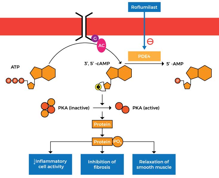
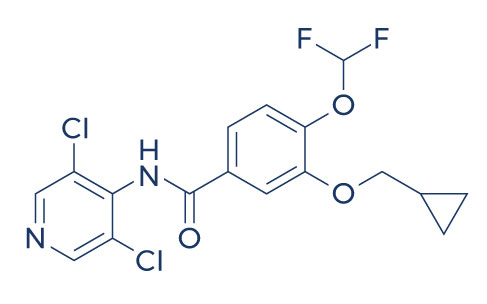
Zoryve (Roflumilast) Cream 0.3%

This medication was approved by the FDA on July 29, 2022 for the treatment of plaque psoriasis in patients aged 12 years and older. It works by inhibiting phosphodiesterase-4 (PDE4), an enzyme responsible for the production of inflammatory substances.

In patients with chronic plaque psoriasis, roflumilast cream administration was safe and highly effective at improving signs and symptoms at doses of 0.5% and 0.15%. [56]

Roflumilast cream administered once daily to affected areas of chronic plaque psoriasis demonstrated superior efficacy to vehicle cream in achieving a state of clear or almost clear at 6 weeks. [57]

In patients with psoriasis vulgaris, topical treatment with a cream formulation of roflumilast resulted in decreased inflammation (as evidenced by a decrease in skin infiltrate thickness) and reduced psoriasis severity. [58]

Non-FDA-Approved Treatment for Psoriasis

KPV (Lysine-Proline-Valine)

KPV is a tripeptide (Lysine-Proline-Valine) that possesses potent anti-inflammatory properties. It’s a C-terminal tripeptide of α-MSH (alpha-Melanocyte-stimulating hormone). Peptides like KPV often act as hormones and relay information from one tissue through the blood to another via biological messengers.

Interestingly, KPV appears to only have an effect in the setting of inflammation and it has almost no effect in normal tissue. The main reason for this is that KPV enters the cells using a transporter that is unregulated in case of inflammation. This suggests that the peptide may serve as an effective or preventive medication in inflammatory conditions like psoriasis. If taken regularly, KPV will be available when necessary and the body can simply excrete it if inflammation and other symptoms are not present.

Once psoriasis begins to resolve, it can leave behind dark spots known as hyperpigmentation. The reason behind this is that psoriasis causes the skin to produce higher levels of inflammatory chemicals. This in turn affects the production of the skin pigment known as melanin, resulting in hyperpigmentation. Another amazing advantage of KPV is that, unlike alpha-Melanocyte-stimulating hormone, it does not cause skin pigmentation. [59] This means that KPV can help treat psoriasis by decreasing inflammation without aggravating hyperpigmentation associated with the disease.

In an imiquimod-induced psoriasis mouse model, the application of a gel containing alpha-MSH resulted in a reduction in psoriasis-associated inflammation. [60] The treatment also significantly decreased psoriatic-like plaque skin sections.

KdPT

KdPT is a tripeptide derivative of α-melanocyte–stimulating hormone (α-MSH or alpha-MSH) which has a similar structure to KPV. Like KPV, KdPT has potent anti-inflammatory effects that can help treat the signs and symptoms of psoriasis.

In murine and human skin affected by psoriasis, KdPT treatment significantly reduced hyperkeratosis (thickening of the outer layer of the skin) and acanthosis (dark skin discoloration in body folds and creases). [61]

In mice with imiquimod-induced psoriasis-like skin lesions, KdPT treatment alleviated the lesions by inhibiting the inflammatory response. [62]

Alpha-MSH was shown to inhibit disease progression in a mouse model of psoriasis-like skin inflammation and prevented the activation and multiplication of effector T cells (attack healthy cells) in subjects with psoriasis. [63]

Bimekizumab

This injectable biologic drug is being tested as a potential treatment for chronic plaque psoriasis. It decreases inflammation by blocking IL-17. Recent studies have found that bimekizumab is safe and effective in patients with psoriasis.

In patients with moderate-to-severe psoriasis, bimekizumab treatment resulted in greater skin clearance over 16 and 48 weeks compared with secukinumab. [64]

Phase 3 trials showed that bimekizumab produced a safety profile similar to the other biologic drugs tested and had higher tolerability. [65]

BE READY, a phase 3 clinical trial, showed that bimekizumab showed high levels of response and was well tolerated by patients with moderate to severe plaque psoriasis. [66]

Janus Kinase Inhibitors (JAK) Inhibitors

Also known as DMARDs (disease-modifying antirheumatic drugs), they are a group of disease-modifying drugs indicated for the treatment of psoriatic arthritis. They work by specifically targeting pathways involved in the production of inflammatory proteins.

In both phase 2 and 3 trials, JAK inhibitors have shown clinical efficacy as measured by the Psoriasis Area and Severity Index 75 response and were associated with higher tolerability. [67]

JAK inhibitors proved to be effective in improving the symptoms of psoriatic arthritis and enhancing the quality of life of patients. [68]

JAK inhibitors were found to have a lower prevalence of side effects compared to other biologic drugs for psoriasis. [69]

6-Thioguanine

This medication is indicated for the treatment of moderate to severe plaque-type psoriasis. It works by stopping the growth of malignant cells by disrupting DNA and RNA.

In patients who have failed to respond to other systemic agents, 6-Thioguanine was found to be an effective treatment. [70]

In patients with moderate to severe plaque-type psoriasis, 6-Thioguanine was found to be safe and effective when administered for defined periods and with careful hematologic monitoring. [71]

In patients with severe recalcitrant psoriasis, 78% of the subjects who received 6-Thioguanine treatment had a dramatic improvement in symptoms. [72]

Azathioprine

This medication is indicated for the treatment of chronic plaque psoriasis. It works by suppressing activated T lymphocyte cells, which in turn stops the production of inflammatory substances.

In patients with chronic plaque psoriasis, azathioprine 300 mg weekly pulse was effective in achieving Psoriasis Area and Severity Index (PASI) of 75 in 42% of the subjects. [73]

Weekly azathioprine pulse appears to be beneficial in treating chronic plaque psoriasis, however, it is less effective than weekly methotrexate. [74]

In patients with psoriasis, azathioprine pulse therapy was associated with a decrease in or disappearance of signs and symptoms of the disease for more than five years. [75]

Fumaric Acid Esters

These are small molecules with immunomodulating, anti-inflammatory, and anti-oxidative properties. They work by reducing the levels of inflammatory substances in the body, making them an effective treatment for moderate to severe psoriasis.

In patients intolerant of or unresponsive to other agents, the use of fumaric acid esters was found to be safe and effective in the management of moderate to severe psoriasis. [76]

In patients with moderate to severe psoriasis, fumaric acid ester treatment was associated with a marked improvement in symptoms after 6 months. [77]

Fumaric acid esters in combination with phototherapy induced a faster therapeutic response with higher tolerability in patients with moderate-to-severe plaque psoriasis. [78]

Mirikizumab

This is a humanized IgG4 monoclonal antibody indicated for the treatment of psoriasis. It works by binding to the inflammatory substance interleukin 23.

Patients treated with mirikizumab 300 mg at 8-week intervals achieved a 90% improvement in the Psoriasis Area and Severity Index (PASI 90) response at week 16. [79]

The administration of mirikizumab in patients with chronic plaque psoriasis was associated with the suppression of inflammatory markers. [80]

Mirikizumab was superior to placebo at week 16 and was associated with higher efficacy and tolerability. [81]

Fluocinonide

This topical steroid helps relieves itching, redness, dryness, crusting, scaling, inflammation, and other discomforts in patients with psoriasis. It works by preventing the release of inflammatory substances in the body.

In patients with refractory psoriasis that had responded poorly to previous therapy, fluocinonide gel (0.05 percent) treatment for 6 weeks completely cleared the lesions. [82]

Fluocinonide treatment was associated with 60-80% clearing of lesions compared to zero with the placebo. [83]

Hydroxyurea

This is an oral cancer medication that is found to be effective in the treatment of psoriasis. It works by reducing the levels of inflammatory molecules.

In patients with psoriasis, the administration of low-dose hydroxyurea was associated with a significant improvement in the Psoriasis Area and Severity Index (PASI) scores. [84]

A study involving patients with chronic plaque psoriasis reported that hydroxyurea is an effective and reasonably safe second-line agent for psoriasis. [85]

In patients with extensive chronic plaque psoriasis, hydroxyurea treatment produced an adequate response as evidenced by a 35% reduction in the Psoriasis Area and Severity Index at or before 8 weeks. [86]

Mycophenolate mofetil

This is an immunosuppressant medication that works by inhibiting activated lymphocytes. As a result, the immune system does not attack healthy skin cells by mistake.

In a man with severe psoriasis, Mycophenolate mofetil improved the psoriasis area and severity index score without short-term side effects. [87]

Mycophenolate mofetil was found to be a good alternative for the treatment of psoriasis in patients who are unable to take methotrexate. [88]

In patients with severe psoriasis, Mycophenolate mofetil was found to be safe and effective. [89]

Sulfasalazine

This is a disease-modifying antirheumatic drug (DMARD) that works by reducing the levels of inflammatory substances.

Sulfasalazine treatment resulted in a significant improvement in inflammatory indices (C-reactive protein and erythrocyte sedimentation rate levels) in patients with active psoriatic arthritis. [90]

The combination of sulfasalazine and pentoxifylline produced a good response in cases of extensive psoriasis. [91]

Sulfasalazine appears to be an effective second-line agent for the treatment of psoriatic arthritis and was found to suppress the immune function by altering B cell number and function. [92]

Tacrolimus

This medication is indicated for eczema and psoriasis. Tacrolimus works by suppressing the immune system to prevent damage to the skin cells.

In patients with facial or intertriginous psoriasis, Tacrolimus treatment was associated with a reduction in disease severity. [93]

In patients with psoriasis on the face and intertriginous areas, Tacrolimus resulted in a significant improvement in the physician’s assessment of the individual signs and symptoms. [94]

Oral tacrolimus has been shown to exert a higher efficacy in the treatment of severe, refractory psoriasis. [95]

Topical Calcineurin Inhibitors

These medications work by altering the immune system, thus, decreasing inflammation associated with psoriasis.

A review of multiple studies demonstrated the efficacy of topical calcineurin inhibitors in the treatment of facial, genital, and intertrigious psoriasis. [96]

A study also found that topical calcineurin inhibitors can significantly reduce facial, genital, and intertriginous psoriatic lesions. [97]

References:

  1. Stein Gold, L., Bagel, J., Allenby, K., & Sidgiddi, S. (2020). Betamethasone dipropionate spray 0.05% alleviates troublesome symptoms of plaque psoriasis. Cutis, 105(2), 97–E1.
  2. Sidgiddi, S., Pakunlu, R. I., & Allenby, K. (2018). Efficacy, Safety, and Potency of Betamethasone Dipropionate Spray 0.05%: A Treatment for Adults with Mild-to-moderate Plaque Psoriasis. The Journal of clinical and aesthetic dermatology, 11(4), 14–22.
  3. Fowler, J. F., Del Rosso, J. Q., Pakunlu, R. I., & Sidgiddi, S. (2017). Treatment Satisfaction, Product Perception, and Quality of Life in Plaque Psoriasis Patients Using Betamethasone Dipropionate Spray 0.05. The Journal of clinical and aesthetic dermatology, 10(11), 13–18.
  4. Guenther, L., Metelitsa, A., & Prajapati, V. H. (2021). Fixed Combination Halobetasol Propionate and Tazarotene Lotion for Plaque Psoriasis. Skin therapy letter, 26(6), 1–3.
  5. Alexis, A. F., Desai, S. R., Han, G., & Jacobson, A. (2021). Fixed-Combination Halobetasol Propionate and Tazarotene Lotion for Psoriasis in Patients With Skin of Color. Journal of drugs in dermatology: JDD, 20(7), 744. https://doi.org/10.36849/JDD.735.
  6. Chung, M., Yeroushalmi, S., Hakimi, M., Bartholomew, E., Liao, W., & Bhutani, T. (2022). A critical review of halobetasol propionate foam (0.05%) as a treatment option for adolescent plaque psoriasis. Expert review of clinical immunology, 1–7. Advance online publication. https://doi.org/10.1080/1744666X.2022.2110071.
  7. Abduelmula, A., Rankin, B. D., Vender, R., Yeung, J., Devani, A. R., & Prajapati, V. H. (2022). Halobetasol Propionate 0.01% Lotion for Plaque Psoriasis and Corticosteroid-Responsive Dermatoses. Skin therapy letter, 27(3), 1–4.
  8. Sugarman, J. L., Weiss, J. S., Tanghetti, E. A., Soung, J., Yamauchi, P. S., Lin, T., Harris, S., Martin, G., & Pillai, R. (2019). Safety and efficacy of halobetasol propionate lotion 0.01% in the treatment of moderate to severe plaque psoriasis: a pooled analysis of 2 phase 3 studies. Cutis, 103(2), 111–116.
  9. Cook-Bolden, F. E., Hebert, A. A., Guenthner, S. T., Kang, R., Martin, G., & Jacobson, A. (2020). Halobetasol Propionate Lotion 0.01% for Moderate-to-Severe Plaque Psoriasis: Pooled Analysis in Male and Female Participants. Journal of drugs in dermatology : JDD, 19(8), 747–754. https://doi.org/10.36849/JDD.2020.5250.
  10. Gottlieb, A. B., Blauvelt, A., Thaçi, D., Leonardi, C. L., Poulin, Y., Drew, J., Peterson, L., Arendt, C., Burge, D., & Reich, K. (2018). Certolizumab pegol for the treatment of chronic plaque psoriasis: Results through 48 weeks from 2 phase 3, multicenter, randomized, double-blinded, placebo-controlled studies (CIMPASI-1 and CIMPASI-2). Journal of the American Academy of Dermatology, 79(2), 302–314.e6. https://doi.org/10.1016/j.jaad.2018.04.012.
  11. Vender, R. B., & Lynde, C. W. (2022). Certolizumab Pegol Use in the Treatment of Moderate-to-Severe Psoriasis: Real-World Data From Two Canadian Centers. Journal of cutaneous medicine and surgery, 26(3), 267–273. https://doi.org/10.1177/12034754221078203.
  12. Lebwohl, M., Blauvelt, A., Paul, C., Sofen, H., Węgłowska, J., Piguet, V., Burge, D., Rolleri, R., Drew, J., Peterson, L., & Augustin, M. (2018). Certolizumab pegol for the treatment of chronic plaque psoriasis: Results through 48 weeks of a phase 3, multicenter, randomized, double-blind, etanercept- and placebo-controlled study (CIMPACT). Journal of the American Academy of Dermatology, 79(2), 266–276.e5. https://doi.org/10.1016/j.jaad.2018.04.013.
  13. Frampton J. E. (2019). Tildrakizumab: A Review in Moderate-to-Severe Plaque Psoriasis. American journal of clinical dermatology, 20(2), 295–306. https://doi.org/10.1007/s40257-019-00435-9.
  14. Näslund-Koch, C., Zachariae, C., & Skov, L. (2020). Tildrakizumab: An Evidence-Based Review of Its Use in the Treatment of Moderate-to-Severe Chronic Plaque Psoriasis. Therapeutics and clinical risk management, 16, 903–916. https://doi.org/10.2147/TCRM.S227880.
  15. Beck, K. M., Sanchez, I. M., Yang, E. J., & Liao, W. (2018). Profile of tildrakizumab-asmn in the treatment of moderate-to-severe plaque psoriasis: evidence to date. Psoriasis (Auckland, N.Z.), 8, 49–58. https://doi.org/10.2147/PTT.S146640.
  16. Howell, S. T., Cardwell, L. A., & Feldman, S. R. (2018). Treating Moderate-to-Severe Plaque Psoriasis With Guselkumab: A Review of Phase II and Phase III Trials. The Annals of pharmacotherapy, 52(4), 380–387. https://doi.org/10.1177/1060028017743268.
  17. Al-Salama, Z. T., & Scott, L. J. (2018). Guselkumab: A Review in Moderate to Severe Plaque Psoriasis. American journal of clinical dermatology, 19(6), 907–918. https://doi.org/10.1007/s40257-018-0406-1.
  18. Nakamura, M., Lee, K., Jeon, C., Sekhon, S., Afifi, L., Yan, D., Lee, K., & Bhutani, T. (2017). Guselkumab for the Treatment of Psoriasis: A Review of Phase III Trials. Dermatology and therapy, 7(3), 281–292. https://doi.org/10.1007/s13555-017-0187-0.
  19. Kokolakis, G., Vadstrup, K., Hansen, J. B., & Carrascosa, J. M. (2022). Brodalumab Is Associated with High Rates of Complete Clearance and Quality of Life Improvement: A Subgroup Analysis of Patients with Psoriasis and Concomitant Psoriatic Arthritis. Dermatology (Basel, Switzerland), 238(4), 620–629. https://doi.org/10.1159/000520290.
  20. Papp, K., Menter, A., Leonardi, C., Soung, J., Weiss, S., Pillai, R., & Jacobson, A. (2020). Long-term efficacy and safety of brodalumab in psoriasis through 120 weeks and after withdrawal and retreatment: subgroup analysis of a randomized phase III trial (AMAGINE-1). The British journal of dermatology, 183(6), 1037–1048. https://doi.org/10.1111/bjd.19132.
  21. Attia, A., Abushouk, A. I., Ahmed, H., Gadelkarim, M., Elgebaly, A., Hassan, Z., Abdel-Daim, M. M., & Negida, A. (2017). Safety and Efficacy of Brodalumab for Moderate-to-Severe Plaque Psoriasis: A Systematic Review and Meta-Analysis. Clinical drug investigation, 37(5), 439–451. https://doi.org/10.1007/s40261-017-0500-9.
  22. Shelton, S. K., Bai, S. R., Jordan, J. K., & Sheehan, A. H. (2019). Ixekizumab: A Review of Its Use for the Management of Moderate to Severe Plaque Psoriasis. The Annals of pharmacotherapy, 53(3), 276–284. https://doi.org/10.1177/1060028018799982.
  23. Farahnik, B., Beroukhim, K., Zhu, T. H., Abrouk, M., Nakamura, M., Singh, R., Lee, K., Bhutani, T., & Koo, J. (2016). Ixekizumab for the Treatment of Psoriasis: A Review of Phase III Trials. Dermatology and therapy, 6(1), 25–37. https://doi.org/10.1007/s13555-016-0102-0.
  24. Syed Y. Y. (2017). Ixekizumab: A Review in Moderate to Severe Plaque Psoriasis. American journal of clinical dermatology, 18(1), 147–158. https://doi.org/10.1007/s40257-017-0254-4.
  25. Menter, A., Tyring, S. K., Gordon, K., Kimball, A. B., Leonardi, C. L., Langley, R. G., Strober, B. E., Kaul, M., Gu, Y., Okun, M., & Papp, K. (2008). Adalimumab therapy for moderate to severe psoriasis: A randomized, controlled phase III trial. Journal of the American Academy of Dermatology, 58(1), 106–115. https://doi.org/10.1016/j.jaad.2007.09.010.
  26. Papoutsaki, M., Chimenti, M. S., Costanzo, A., Talamonti, M., Zangrilli, A., Giunta, A., Bianchi, L., & Chimenti, S. (2007). Adalimumab for severe psoriasis and psoriatic arthritis: an open-label study in 30 patients previously treated with other biologics. Journal of the American Academy of Dermatology, 57(2), 269–275. https://doi.org/10.1016/j.jaad.2006.12.003.
  27. Pitarch, G., Sanchez-Carazo, J. L., Mahiques, L., Perez-Ferriols, M. A., & Fortea, J. M. (2007). Treatment of psoriasis with adalimumab. Clinical and experimental dermatology, 32(1), 18–22. https://doi.org/10.1111/j.1365-2230.2006.02288.x.
  28. Leonardi, C. L., Powers, J. L., Matheson, R. T., Goffe, B. S., Zitnik, R., Wang, A., Gottlieb, A. B., & Etanercept Psoriasis Study Group (2003). Etanercept as monotherapy in patients with psoriasis. The New England journal of medicine, 349(21), 2014–2022. https://doi.org/10.1056/NEJMoa030409.
  29. Iyer, S., Yamauchi, P., & Lowe, N. J. (2002). Etanercept for severe psoriasis and psoriatic arthritis: observations on combination therapy. The British journal of dermatology, 146(1), 118–121. https://doi.org/10.1046/j.0007-0963.2001.04529.x.
  30. Kokelj, F., Miertusova Tothova, S., Patamia, M., & Trevisan, G. (2006). Our experience with etanercept in the treatment of psoriasis. Acta dermatovenerologica Croatica : ADC, 14(4), 241–245.
  31. Antoniou, C., Stefanaki, I., Stratigos, A., Moustou, E., Vergou, T., Stavropoulos, P., Avgerinou, G., Rigopoulos, D., & Katsambas, A. D. (2010). Infliximab for the treatment of psoriasis in Greece: 4 years of clinical experience at a single centre. The British journal of dermatology, 162(5), 1117–1123. https://doi.org/10.1111/j.1365-2133.2009.09578.x
  32. Cassano, N., Loconsole, F., Amoruso, A., Coviello, C., Filieri, M., Filotico, R., Del Vecchio, S., & Vena, G. A. (2004). Infliximab monotherapy for refractory psoriasis: preliminary results. International journal of immunopathology and pharmacology, 17(3), 373–380. https://doi.org/10.1177/039463200401700317.
  33. Reich, K., Nestle, F. O., Papp, K., Ortonne, J. P., Evans, R., Guzzo, C., Li, S., Dooley, L. T., Griffiths, C. E., & EXPRESS study investigators (2005). Infliximab induction and maintenance therapy for moderate-to-severe psoriasis: a phase III, multicentre, double-blind trial. Lancet (London, England), 366(9494), 1367–1374. https://doi.org/10.1016/S0140-6736(05)67566-6.
  34. Patel, N. U., Felix, K., Reimer, D., & Feldman, S. R. (2017). Calcipotriene/betamethasone dipropionate for the treatment of psoriasis vulgaris: an evidence-based review. Clinical, cosmetic and investigational dermatology, 10, 385–391. https://doi.org/10.2147/CCID.S131727.
  35. White, S., Vender, R., Thaçi, D., Haverkamp, C., Naeyaert, J. M., Foster, R., Martinez Escribano, J. A., Cambazard, F., & Bibby, A. (2006). Use of calcipotriene cream (Dovonex cream) following acute treatment of psoriasis vulgaris with the calcipotriene/betamethasone dipropionate two-compound product (Taclonex): a randomized, parallel-group clinical trial. American journal of clinical dermatology, 7(3), 177–184. https://doi.org/10.2165/00128071-200607030-00004.
  36. Rogalski C. (2015). Calcipotriol/betamethasone for the treatment of psoriasis: efficacy, safety, and patient acceptability. Psoriasis (Auckland, N.Z.), 5, 97–107. https://doi.org/10.2147/PTT.S63127.
  37. Narbutt, J., Czajkowski, R., Lesiak, A., Owczarek, W., Reich, A., & Szepietowski, J. C. (2021). Calcipotriene/betamethasone dipropionate foam: primary evidence supporting its use in patients with psoriasis vulgaris. A narrative review. Postepy dermatologii i alergologii, 38(5), 727–733. https://doi.org/10.5114/ada.2021.110062.
  38. Papp, K. A., Guenther, L., Boyden, B., Larsen, F. G., Harvima, R. J., Guilhou, J. J., Kaufmann, R., Rogers, S., van de Kerkhof, P. C., Hanssen, L. I., Tegner, E., Burg, G., Talbot, D., & Chu, A. (2003). Early onset of action and efficacy of a combination of calcipotriene and betamethasone dipropionate in the treatment of psoriasis. Journal of the American Academy of Dermatology, 48(1), 48–54. https://doi.org/10.1067/mjd.2003.130.
  39. Feldman, S. R., Levi, E., Pathak, P., Kakatkar, S., & Balkrishnan, R. (2013). Using a single product (calcipotriene/betamethasone topical suspension) vs multiple products to manage body and scalp psoriasis: comparisons in resource utilization and costs. Journal of medical economics, 16(12), 1405–1413. https://doi.org/10.3111/13696998.2013.848209.
  40. Jemec, G. B., van de Kerkhof, P. C., Enevold, A., & Ganslandt, C. (2011). Significant one week efficacy of a calcipotriol plus betamethasone dipropionate scalp formulation. Journal of the European Academy of Dermatology and Venereology : JEADV, 25(1), 27–32. https://doi.org/10.1111/j.1468-3083.2010.03682.x.
  41. Hashim, P. W., Nia, J. K., Terrano, D., Goldenberg, G., & Kircik, L. H. (2017). A Comparative Study to Evaluate Epidermal Barrier Integrity of Psoriasis Patients Treated With Calcipotriene/Betamethasone Topical Suspension Versus Betamethasone Dipropionate 0.05% Lotion. Journal of drugs in dermatology : JDD, 16(8), 747–752.
  42. Shepherd, J., Taheri, A., & Feldman, S. R. (2013). Once-daily topical treatment for psoriasis: calcipotriene + betamethasone two-compound topical formulation. Clinical, cosmetic and investigational dermatology, 7, 19–22. https://doi.org/10.2147/CCID.S56673.
  43. White, S., Vender, R., Thaçi, D., Haverkamp, C., Naeyaert, J. M., Foster, R., Martinez Escribano, J. A., Cambazard, F., & Bibby, A. (2006). Use of calcipotriene cream (Dovonex cream) following acute treatment of psoriasis vulgaris with the calcipotriene/betamethasone dipropionate two-compound product (Taclonex): a randomized, parallel-group clinical trial. American journal of clinical dermatology, 7(3), 177–184. https://doi.org/10.2165/00128071-200607030-00004.
  44. Augustin, M., Peeters, P., Radtke, M., Moehling, U., & Lapp, C. (2007). Cost-effectiveness model of topical treatment of mild to moderate psoriasis vulgaris in Germany. A comparison of calcipotriol/betamethasone (Daivobet/Dovobet/Taclonex) once daily and a morning/evening non-fix combination of calcipotriol and betamethasone. Dermatology (Basel, Switzerland), 215(3), 219–228. https://doi.org/10.1159/000106791.
  45. Highton, A., & Quell, J. (1995). Calcipotriene ointment 0.005% for psoriasis: a safety and efficacy study. Calcipotriene Study Group. Journal of the American Academy of Dermatology, 32(1), 67–72. https://doi.org/10.1016/0190-9622(95)90186-8.
  46. Gao, J. C., Emmerich, V. K., Feldman, S., & Strowd, L. C. (2021). Evaluating calcipotriene 0.005% foam for the treatment of plaque psoriasis of the scalp and body in patients aged 12 years and older. Expert opinion on pharmacotherapy, 22(5), 543–547. https://doi.org/10.1080/14656566.2020.1853099.
  47. Scott, L. J., Dunn, C. J., & Goa, K. L. (2001). Calcipotriol ointment. A review of its use in the management of psoriasis. American journal of clinical dermatology, 2(2), 95–120. https://doi.org/10.2165/00128071-200102020-00008.
  48. Feldman, S. R., Matheson, R., Bruce, S., Grande, K., Markowitz, O., Kempers, S., Brundage, T., Wyres, M., & U0267-301 & 302 Study Investigators (2012). Efficacy and safety of calcipotriene 0.005% foam for the treatment of plaque-type psoriasis: results of two multicenter, randomized, double-blind, vehicle-controlled, phase III clinical trials. American journal of clinical dermatology, 13(4), 261–271. https://doi.org/10.2165/11630710-000000000-00000.
  49. Feldman, S. R., Mills, M., Brundage, T., & Eastman, W. J. (2013). A multicenter, randomized, double-blind study of the efficacy and safety of calcipotriene foam, 0.005%, vs vehicle foam in the treatment of plaque-type psoriasis of the scalp. Journal of drugs in dermatology : JDD, 12(3), 300–306.
  50. Blair H. A. (2020). Risankizumab: A Review in Moderate to Severe Plaque Psoriasis. Drugs, 80(12), 1235–1245. https://doi.org/10.1007/s40265-020-01357-1.
  51. Kristensen, L. E., Keiserman, M., Papp, K., McCasland, L., White, D., Lu, W., Wang, Z., Soliman, A. M., Eldred, A., Barcomb, L., & Behrens, F. (2022). Efficacy and safety of risankizumab for active psoriatic arthritis: 24-week results from the randomised, double-blind, phase 3 KEEPsAKE 1 trial. Annals of the rheumatic diseases, 81(2), 225–231. https://doi.org/10.1136/annrheumdis-2021-221019.
  52. Reddy, V., Yang, E. J., Myers, B., & Liao, W. (2020). Clinical Evaluation of Risankizumab-rzaa in the Treatment of Plaque Psoriasis. Journal of inflammation research, 13, 53–60. https://doi.org/10.2147/JIR.S215196.
  53. Weber, J., & Keam, S. J. (2009). Ustekinumab. BioDrugs : clinical immunotherapeutics, biopharmaceuticals and gene therapy, 23(1), 53–61. https://doi.org/10.2165/00063030-200923010-00006.
  54. Zaghi, D., Krueger, G. G., & Callis Duffin, K. (2012). Ustekinumab: a review in the treatment of plaque psoriasis and psoriatic arthritis. Journal of drugs in dermatology : JDD, 11(2), 160–167.
  55. Philipp, S., Menter, A., Nikkels, A. F., Barber, K., Landells, I., Eichenfield, L. F., Song, M., Randazzo, B., Li, S., Hsu, M. C., Zhu, Y., DePrimo, S., & Paller, A. S. (2020). Ustekinumab for the treatment of moderate-to-severe plaque psoriasis in paediatric patients (≥ 6 to < 12 years of age): efficacy, safety, pharmacokinetic and biomarker results from the open-label CADMUS Jr study. The British journal of dermatology, 183(4), 664–672. https://doi.org/10.1111/bjd.19018.
  56. Papp, K. A., Gooderham, M., Droege, M., Merritt, C., Osborne, D. W., Berk, D. R., Thurston, A. W., Smith, V. H., & Welgus, H. (2020). Roflumilast Cream Improves Signs and Symptoms of Plaque Psoriasis: Results from a Phase 1/2a Randomized, Controlled Study. Journal of drugs in dermatology: JDD, 19(8), 734–740. https://doi.org/10.36849/JDD.2020.5370.
  57. Lebwohl, M. G., Papp, K. A., Stein Gold, L., Gooderham, M. J., Kircik, L. H., Draelos, Z. D., Kempers, S. E., Zirwas, M., Smith, K., Osborne, D. W., Trotman, M. L., Navale, L., Merritt, C., Berk, D. R., Welgus, H., & ARQ-151 201 Study Investigators (2020). Trial of Roflumilast Cream for Chronic Plaque Psoriasis. The New England journal of medicine, 383(3), 229–239. https://doi.org/10.1056/NEJMoa2000073.
  58. Snape, S. D., Wigger-Alberti, W., & Goehring, U. M. (2016). A phase I randomized trial to assess the effect on skin infiltrate thickness and tolerability of topical phosphodiesterase inhibitors in the treatment of psoriasis vulgaris using a modified psoriasis plaque test. The British journal of dermatology, 175(3), 479–486. https://doi.org/10.1111/bjd.14634.
  59. Brzoska, T., Luger, T. A., Maaser, C., Abels, C., & Böhm, M. (2008). Alpha-melanocyte-stimulating hormone and related tripeptides: biochemistry, antiinflammatory and protective effects in vitro and in vivo, and future perspectives for the treatment of immune-mediated inflammatory diseases. Endocrine reviews, 29(5), 581–602. https://doi.org/10.1210/er.2007-0027.
  60. Available from https://www.researchgate.net/publication/284175171_Percutaneous_delivery_of_a_-melanocyte-stimulating_hormone_for_the_treatment_of_imiquimod-induced_psoriasis.
  61. Mykicki, N., Klenner, L., Baumann, C., Auriemma, M., Sternemann, C., Soeberdt, M., Elliott, G. R., Abels, C., Luger, T. A., & Loser, K. (2017). The tripeptide KdPT ameliorates ongoing psoriasis-like skin inflammation in murine and human skin. Experimental dermatology, 26(4), 328–334. https://doi.org/10.1111/exd.13145.
  62. Tang, J., Zhou, Z., Qian, J., Lin, X., Liu, Q., Xie, Q., & Xiong, S. (2022). KdPT alleviates imiquimod-induced psoriasis-like skin lesion in mice via inhibiting proliferation and inflammation response. Die Pharmazie, 77(2), 48–53. https://doi.org/10.1691/ph.2022.1970.
  63. Auriemma, M., Brzoska, T., Klenner, L., Kupas, V., Goerge, T., Voskort, M., Zhao, Z., Sparwasser, T., Luger, T. A., & Loser, K. (2012). α-MSH-stimulated tolerogenic dendritic cells induce functional regulatory T cells and ameliorate ongoing skin inflammation. The Journal of investigative dermatology, 132(7), 1814–1824. https://doi.org/10.1038/jid.2012.59.
  64. Reich, K., Warren, R. B., Lebwohl, M., Gooderham, M., Strober, B., Langley, R. G., Paul, C., De Cuyper, D., Vanvoorden, V., Madden, C., Cioffi, C., Peterson, L., & Blauvelt, A. (2021). Bimekizumab versus Secukinumab in Plaque Psoriasis. The New England journal of medicine, 385(2), 142–152. https://doi.org/10.1056/NEJMoa2102383.
  65. Freitas, E., Blauvelt, A., & Torres, T. (2021). Bimekizumab for the Treatment of Psoriasis. Drugs, 81(15), 1751–1762. https://doi.org/10.1007/s40265-021-01612-z.
  66. Ruggiero, A., Potestio, L., Camela, E., Fabbrocini, G., & Megna, M. (2022). Bimekizumab for the Treatment of Psoriasis: A Review of the Current Knowledge. Psoriasis (Auckland, N.Z.), 12, 127–137. https://doi.org/10.2147/PTT.S367744.
  67. Kvist-Hansen, A., Hansen, P. R., & Skov, L. (2020). Systemic Treatment of Psoriasis with JAK Inhibitors: A Review. Dermatology and therapy, 10(1), 29–42. https://doi.org/10.1007/s13555-019-00347-w.
  68. Wang, L., Ping, X., Chen, W., & Xing, W. (2021). Performance of Janus kinase inhibitors in psoriatic arthritis with axial involvement in indirect comparison with ankylosing spondylitis: a retrospective analysis from pooled data. Clinical rheumatology, 40(5), 1725–1737. https://doi.org/10.1007/s10067-020-05442-4.
  69. Słuczanowska-Głąbowska, S., Ziegler-Krawczyk, A., Szumilas, K., & Pawlik, A. (2021). Role of Janus Kinase Inhibitors in Therapy of Psoriasis. Journal of clinical medicine, 10(19), 4307. https://doi.org/10.3390/jcm10194307.
  70. Zackheim, H. S., Glogau, R. G., Fisher, D. A., & Maibach, H. I. (1994). 6-Thioguanine treatment of psoriasis: experience in 81 patients. Journal of the American Academy of Dermatology, 30(3), 452–458. https://doi.org/10.1016/s0190-9622(94)70055-9.
  71. Murphy, F. P., Coven, T. R., Burack, L. H., Gilleaudeau, P., Cardinale, I., Auerbach, R., & Krueger, J. G. (1999). Clinical clearing of psoriasis by 6-thioguanine correlates with cutaneous T-cell depletion via apoptosis: evidence for selective effects on activated T lymphocytes. Archives of dermatology, 135(12), 1495–1502. https://doi.org/10.1001/archderm.135.12.1495.
  72. Mason, C., & Krueger, G. G. (2001). Thioguanine for refractory psoriasis: a 4-year experience. Journal of the American Academy of Dermatology, 44(1), 67–72. https://doi.org/10.1067/mjd.2001.109296.
  73. Verma, P., Verma, K. K., Khanna, N., Gupta, S., & Bhari, N. (2016). Effectiveness of weekly azathioprine pulse in the treatment of chronic plaque psoriasis: an open-label study. Clinical and experimental dermatology, 41(7), 717–722. https://doi.org/10.1111/ced.12887.
  74. Verma, K. K., Kumar, P., Bhari, N., Gupta, S., & Kalaivani, M. (2021). Azathioprine weekly pulse versus methotrexate for the treatment of chronic plaque psoriasis: A randomized controlled trial. Indian journal of dermatology, venereology and leprology, 87(4), 509–514. https://doi.org/10.25259/IJDVL_718_18.
  75. Gupta R. (2015). Prolonged Remission of Psoriasis with Azathioprine Pulse Therapy. Indian journal of dermatology, 60(4), 360–363. https://doi.org/10.4103/0019-5154.160480.
  76. Smith D. (2017). Fumaric acid esters for psoriasis: a systematic review. Irish journal of medical science, 186(1), 161–177. https://doi.org/10.1007/s11845-016-1470-2.
  77. Mrowietz, U., Christophers, E., & Altmeyer, P. (1998). Treatment of psoriasis with fumaric acid esters: results of a prospective multicentre study. German Multicentre Study. The British journal of dermatology, 138(3), 456–460. https://doi.org/10.1046/j.1365-2133.1998.02124.x.
  78. Weisenseel, P., Reich, K., Griemberg, W., Merten, K., Gröschel, C., Gomez, N. N., Taipale, K., Bräu, B., & Zschocke, I. (2017). Efficacy and safety of fumaric acid esters in combination with phototherapy in patients with moderate-to-severe plaque psoriasis (FAST). Journal der Deutschen Dermatologischen Gesellschaft = Journal of the German Society of Dermatology : JDDG, 15(2), 180–186. https://doi.org/10.1111/ddg.12837.
  79. Reich, K., Rich, P., Maari, C., Bissonnette, R., Leonardi, C., Menter, A., Igarashi, A., Klekotka, P., Patel, D., Li, J., Tuttle, J., Morgan-Cox, M., Edson-Heredia, E., Friedrich, S., Papp, K., & AMAF investigators (2019). Efficacy and safety of mirikizumab (LY3074828) in the treatment of moderate-to-severe plaque psoriasis: results from a randomized phase II study. The British journal of dermatology, 181(1), 88–95. https://doi.org/10.1111/bjd.17628.
  80. Salimi, S., Yamauchi, P. S., Thakur, R., Weinberg, J. M., Kircik, L., Abdelmaksoud, A., Wollina, U., Lotti, T., Sharma, A., Grabbe, S., & Goldust, M. (2020). Interleukin 23p19 inhibitors in chronic plaque psoriasis with focus on mirikizumab: A narrative review. Dermatologic therapy, 33(4), e13800. https://doi.org/10.1111/dth.13800.
  81. Blauvelt, A., Kimball, A. B., Augustin, M., Okubo, Y., Witte, M. M., Capriles, C. R., Sontag, A., Arora, V., Osuntokun, O., & Strober, B. (2022). Efficacy and safety of mirikizumab in psoriasis: results from a 52-week, double-blind, placebo-controlled, randomized withdrawal, phase III trial (OASIS-1). The British journal of dermatology, 10.1111/bjd.21743. Advance online publication. https://doi.org/10.1111/bjd.21743.
  82. Willoughby, C. B., & Cook, W. R., Jr (1977). Treatment of refractory psoriasis with fluocinonide gel. Cutis, 20(1), 144–145.
  83. Lee, C. S., & Koo, J. (2009). The efficacy of three class I topical synthetic corticosteroids, fluocinonide 0.1% cream, clobetasol 0.05% cream and halobetasol 0.05% cream: a Scholtz-Dumas bioassay comparison. Journal of drugs in dermatology : JDD, 8(8), 751–755.
  84. Mahbub, M. S., Khondker, L., Khan, S. I., & Hazra, S. C. (2013). Comparative efficacy of hydroxyurea and methotrexate in treating psoriasis. Mymensingh medical journal : MMJ, 22(1), 116–130.
  85. Sharma, V. K., Dutta, B., & Ramam, M. (2004). Hydroxyurea as an alternative therapy for psoriasis. Indian journal of dermatology, venereology and leprology, 70(1), 13–17.
  86. Kumar, B., Saraswat, A., & Kaur, I. (2001). Rediscovering hydroxyurea: its role in recalcitrant psoriasis. International journal of dermatology, 40(8), 530–534. https://doi.org/10.1046/j.1365-4362.2001.01255.x
  87. Haufs, M. G., Beissert, S., Grabbe, S., Schütte, B., & Luger, T. A. (1998). Psoriasis vulgaris treated successfully with mycophenolate mofetil. The British journal of dermatology, 138(1), 179–181. https://doi.org/10.1046/j.1365-2133.1998.02048.x.
  88. Akhyani, M., Chams-Davatchi, C., Hemami, M. R., & Fateh, S. (2010). Efficacy and safety of mycophenolate mofetil vs. methotrexate for the treatment of chronic plaque psoriasis. Journal of the European Academy of Dermatology and Venereology : JEADV, 24(12), 1447–1451. https://doi.org/10.1111/j.1468-3083.2010.03667.x.
  89. Geilen, C. C., Arnold, M., & Orfanos, C. E. (2001). Mycophenolate mofetil as a systemic antipsoriatic agent: positive experience in 11 patients. The British journal of dermatology, 144(3), 583–586. https://doi.org/10.1046/j.1365-2133.2001.04088.x.
  90. Farr, M., Kitas, G. D., Waterhouse, L., Jubb, R., Felix-Davies, D., & Bacon, P. A. (1988). Treatment of psoriatic arthritis with sulphasalazine: a one year open study. Clinical rheumatology, 7(3), 372–377. https://doi.org/10.1007/BF02239195.
  91. el-Mofty, M., el-Darouti, M., Rasheed, H., Bassiouny, D. A., Abdel-Halim, M., Zaki, N. S., el-Hanafy, G., el-Hadidi, H., Azzam, O., el-Ramly, A., & Fawzy, M. (2011). Sulfasalazine and pentoxifylline in psoriasis: a possible safe alternative. The Journal of dermatological treatment, 22(1), 31–37. https://doi.org/10.3109/09546630903460260.
  92. Newman, E. D., Perruquet, J. L., & Harrington, T. M. (1991). Sulfasalazine therapy in psoriatic arthritis: clinical and immunologic response. The Journal of rheumatology, 18(9), 1379–1382.
  93. Lebwohl, M., Freeman, A. K., Chapman, M. S., Feldman, S. R., Hartle, J. E., Henning, A., & Tacrolimus Ointment Study Group (2004). Tacrolimus ointment is effective for facial and intertriginous psoriasis. Journal of the American Academy of Dermatology, 51(5), 723–730. https://doi.org/10.1016/j.jaad.2004.07.011.
  94. Freeman, A. K., Linowski, G. J., Brady, C., Lind, L., Vanveldhuisen, P., Singer, G., & Lebwohl, M. (2003). Tacrolimus ointment for the treatment of psoriasis on the face and intertriginous areas. Journal of the American Academy of Dermatology, 48(4), 564–568. https://doi.org/10.1067/mjd.2003.169.
  95. Malecic, N., & Young, H. (2016). Tacrolimus for the management of psoriasis: clinical utility and place in therapy. Psoriasis (Auckland, N.Z.), 6, 153–163. https://doi.org/10.2147/PTT.S101233.
  96. Wang, C., & Lin, A. (2014). Efficacy of topical calcineurin inhibitors in psoriasis. Journal of cutaneous medicine and surgery, 18(1), 8–14. https://doi.org/10.2310/7750.2013.13059.
  97. Dattola, A., Silvestri, M., Bennardo, L., Del Duca, E., Longo, C., Bianchi, L., & Nisticò, S. (2018). Update of calcineurin inhibitors to treat inverse psoriasis: A systematic review. Dermatologic therapy, 31(6), e12728. https://doi.org/10.1111/dth.12728.

Methylation

The term methylation refers to the biochemical process that is utilized in your body for the transport of essential nutrients, production of energy, and modulation of genes. With decreased methylation activity, also known as methylation deficiency, your body’s ability to perform various important chemical functions becomes impaired. This in turn can increase your risk of developing a broad range of medical conditions such as seizures, migraines, dizziness, mood problems, mental disorders, infertility, infections, autoimmune disease, and pregnancy complications. To keep your body working at optimum levels, you need appropriate supplementation with the proper methylated vitamins and nutrients.

The Methylation Cycle

The proper functioning of the methylation cycle ensures that the critical reactions in your body are maintained. Methylation    occurs  in every cell of your body over 250 billion times per second. This process is simply the addition of a small methyl group to a substrate (the substance on which an enzyme acts). Methylation leads to the activation of the methylene tetrahydrofolate reductase (MTHFR) enzyme. MTHFR is responsible for the conversion of dietary folate to activated folate – a process that occurs in every cell of your body and is vital for the maintenance of optimal health. MTHFR is not the only vital enzyme in the methylation cycle. There are also many other enzymes. The inefficiency of MTHFR and other important enzymes can lead to a wide array of medical conditions. Currently, these genetic alterations can be identified by DNA analysis to help determine the root cause of your symptoms.

Aside from gene activation, methylation is also responsible for controlling inflammation via regulation of homocysteine levels, building neurotransmitters (brain chemicals), liver detoxification of harmful substances via regulation of glutathione levels, building immune cells, producing energy in your cells, DNA   and      RNA            synthesis, processing hormones, and producing protective covering of nerves.

MTHFR Mutation

MTHFR is a gene that gives your body instructions for producing the methylene tetrahydrofolate reductase enzyme. In some cases, there can be one or more abnormal variants of this gene that can be passed down from the parent to the child. In general, the more variations you have, the higher your risk of developing health issues.  A MTHFR gene mutation can have a negative impact on your overall health by changing the way your body metabolizes and convert dietary nutrients. As a result, these nutrients cannot be effectively converted into active vitamins, minerals, and proteins. This in turn affects your overall energy production. In addition, MTHFR gene mutation can have a detrimental effect on the levels of neurotransmitters and bodily functions such as cognition, digestion, and other vital processes.

Symptoms of Poor Methylation

This can lead to unpleasant symptoms, such as:

  • Addictions
  • Allergies
  • Anxiety
  • Depression
  • Digestive problems
  • Fatigue and low energy levels
  • Headaches and migraines
  • Insomnia (sleeping difficulties)
  • Multiple miscarriages
  • Muscle pain

Health Issues associated with Poor Methylation

MTHFR gene mutations can lead to poor methylation. This in turn causes a broad range of health issues. They can vary in severity as they affect everyone differently.

Attention Deficit Hyperactivity Disorder (ADHD)

ADHD is a condition that affects attention and self-control. Studies suggest that impaired methylation results in inadequate production of the neurotransmitter known as dopamine. [1-7] Dopamine plays a role in motor control, reward, motivation, and cognitive function.

Allergies

Evidence suggests that impaired methylation can lead to various allergies as it can cause imbalanced TH1/TH2 ratio and high histamine levels. [8-11] This increased immune response stimulates the activation and recruitment of immune cells (e.g. TH1, TH2, IgE antibodies, or eosinophils) which in turn results in allergic inflammation.

Autism

This complex, lifelong developmental disability is characterized by impairment in social skills, communication, relationships, and self-regulation. A good deal of evidence suggests that people with autism have abnormal blood levels of methionine (Met), S-adenosylmethionine (SAM), S-adenosylhomocysteine (SAH), and the SAM/SAH ratio – these compounds are considered predictive indices of cellular methylation ability. [12-17]

Cancer

Studies suggest that abnormal methylation (both overmethylation and undermethylation) can lead to the development of various types of cancer such as gastrointestinal, brain, head, and neck cancer. [18- 26] The mechanism behind this is that impaired methylation leads to a state of immune surveillance dysfunction. [27-30] As a result, the immune system is less likely to recognize abnormal cell surface antigens on precancerous cells.

Increased Body Toxins

Studies suggest that alterations in methylation impair the body’s ability to excrete toxins by decreasing the body’s glutathione stores. [31-35] Glutathione is one of the major antioxidants and is responsible for fighting free radicals. It is also helps maintain the levels of the antioxidants vitamins C and E, and detoxify heavy metals and other foreign molecules in the body.

Low Energy Levels/Chronic Fatigue

Methylation is vital for energy production within the cells. It does this by supporting the production of coenzyme Q10 (CoQ10) and mitochondrial fatty acid oxidation. According to studies, impaired methylation can lead to low energy levels, chronic fatigue syndrome, and chronic pain syndromes. [36-41]

Cardiovascular Disease

Impaired methylation inhibits the conversion from the molecule homocysteine to methionine and leads to dysfunction in the blood vessels of the heart – both of which significantly increases the risk of cardiovascular disease. [42-50] In addition, breakdowns within the cycle decrease the level of CoQ10 which in turn increases the risk of congestive heart failure and other heart diseases. [51-53]

Mood Problems/Psychiatric Disorders

The methylation cycle is important for the production of various neurotransmitters such as dopamine, serotonin, melatonin, epinephrine, and norepinephrine. Impairment in this cycle increases the risk of depression, anxiety, schizophrenia, bipolar disorder, and other serious mental illnesses. [54-62] This is because neurotransmitters help regulate mood, appetite, sleep pattern, and the body’s circadian rhythm.

Birth Defects

Methylation involves the conversion of dietary folate to activated folate. Folate is very important during pregnancy as it helps in red blood cell formation and maintenance of healthy cell growth and function. Impaired methylation can lead to folate deficiency which in turn increases the risk of birth defects of the brain and spine. [63-70]

Obesity/Unhealthy Weight

The methylation cycle is also vital for maintaining a healthy weight by promoting effective fat metabolism. Studies found a strong link between impaired methylation and increased risk of obesity and being overweight. [71-76]

Faster Aging

Any impairment in the methylation cycle can speed up the aging process by affecting numerous body systems. In addition, it can lead to many age-related degenerative processes as well as increased mortality rate. [77-83]

Weak Immune System

Methylation results in the DNA needed for the production of new cells of the immune system. New T cells are integral part of the immune system’s response to infection and other foreign bodies. Poor methylation ultimately leads to poor regulatory control of immune cells which significantly increases the risk of infection and autoimmune disease. [84-91]

Demyelination Disorders

Myelination refers to the coating of neurons with myelin sheath, which is known as the protective covering of neurons. This myelin sheath allows electrical impulses in the brain and spinal cord to be transmitted at a faster and efficient manner. Impaired methylation affects the process of myelination which leads to damage to neurons, resulting in demyelination disorders such as multiple sclerosis. [92-100]

Hormonal Imbalance

Methylation plays an important role in metabolizing and detoxifying estrogen, a hormone that promotes the development and maintenance of female body characteristics. With impaired methylation, the process of estrogen metabolism and detoxification is slow, resulting in premenstrual syndrome, polycystic ovarian syndrome, fibroids, endometriosis, and heavy menstruation. [101-107]

Hypertension

Methylation and blood pressure are strongly linked. A convincing number of studies found that patients with higher systolic blood pressure, diastolic blood pressure, and/or hypertension have lower methylation levels. [108-117]

Male Infertility

Impaired methylation is also known to cause infertility in men. An overwhelming body of clinical evidence suggests that abnormal DNA methylation is associated with sperm alterations such as impaired sperm production and low sperm count, resulting in infertility. [118-127]

Cognitive Dysfunction

Memory, learning, and other cognitive functions can also be affected by abnormal methylation. A number of high-quality studies have linked poor methylation to cognitive problems such as dementia, Parkinson’s disease, and Alzheimer’s disease. [128-141]

Digestive Problems

Effective methylation is essential for the production of bile, a thick, yellow-green fluid that is produced by the liver and is stored in the gallbladder. This fluid has anti-microbial properties and aids in the absorption of vitamin A, D, E and K. In addition, bile helps excrete toxins and excess cholesterol. Studies show that poor methylation leads to inadequate phosphatidylcholine, a key component of bile, resulting in digestive problems such as fat malabsorption and gall stones. [142-148]

Inflammatory Disorders

When the availability of methylated molecules in the body is negatively affected, various essential processes will be impaired. As a result, the levels of inflammatory substances start to increase, leading to the development of different inflammatory disorders such as arthritis and inflammatory bowel disease (IBD). [149-156]

Treatment for Poor Methylation

A methyl group (CH3) is provided to your body through a universal methyl donor called SAMe (S-adenosylmethionine). In this way, methylation occurs which enables different body systems to perform at optimal levels. In order for the system that produces SAMe to function at a hundred percent, it requires 5-MTHF, the most active form of folate, or vitamin B9. With adequate amounts of 5-MTHF, the methylation cycle will work efficiently.

To enhance the methylation process, SAMe levels should be restored. There are several ways of increasing SAMe levels such as taking supplements that contain the following:

  • B12 (Methylcobalamin / Hydroxycobalamin)
  • SAM E, 5 methyltetrahydrofolate
  • TMG (trimethylglycine)
  • Vitamin B-6 (pyridoxal 5’-phosphate)
  • Vitamin B2 (riboflavin)
  • Quatrefolic
  • 5-Methyltetrahydrofolate
  • Choline
  • Methionine
  • Polyphenols
  • Genistein
  • Magnesium
  • Vitamin D
  • Calcium (as dicalcium phosphate)

To find out if you have a problem with your methylation cycle, you can undergo a simple genetic test. This test will analyse your blood or saliva to determine the presence of genetic changes that may affect important biochemical processes in your body. After the test, a treatment plan specific to your needs will be created to improve or restore your methylation cycle.

References:

  1. Tonelli E, Pascale E, Troianiello M, D’Addario C, Adriani W. DAT1 Gene Methylation as an Epigenetic Biomarker in Attention Deficit Hyperactivity Disorder: A Commentary. Front Genet. 2020;11:444. Published 2020 May 11. doi:10.3389/fgene.2020.00444.
  2. Heinrich H, Grunitz J, Stonawski V, et al. Attention, cognitive control and motivation in ADHD: Linking event-related brain potentials and DNA methylation patterns in boys at early school age. Sci Rep. 2017;7(1):3823. Published 2017 Jun 19. doi:10.1038/s41598-017-03326-3.
  3. Hodgson NW, Waly MI, Trivedi MS, Power-Charnitsky VA, Deth RC. Methylation-related metabolic effects of D4 dopamine receptor expression and activation. Transl Psychiatry. 2019;9(1):295. Published 2019 Nov 12. doi:10.1038/s41398-019-0630-3.
  4. Neumann A, Walton E, Alemany S, et al. Association between DNA methylation and ADHD symptoms from birth to school age: a prospective meta-analysis. Transl Psychiatry. 2020;10(1):398. Published 2020 Nov 12. doi:10.1038/s41398-020-01058-z.
  5. Weiß, A. L., Meijer, M., Budeus, B., Pauper, M., Hakobjan, M., Groothuismink, J., Shi, Y., Neveling, K., Buitelaar, J. K., Hoogman, M., Franke, B., & Klein, M. (2021). DNA methylation associated with persistent ADHD suggests TARBP1 as novel candidate. Neuropharmacology, 184, 108370. https://doi.org/10.1016/j.neuropharm.2020.108370.
  6. Wilmot, B., Fry, R., Smeester, L., Musser, E. D., Mill, J., & Nigg, J. T. (2016). Methylomic analysis of salivary DNA in childhood ADHD identifies altered DNA methylation in VIPR2. Journal of child psychology and psychiatry, and allied disciplines, 57(2), 152–160. https://doi.org/10.1111/jcpp.12457.
  7. Mooney MA, Ryabinin P, Wilmot B, Bhatt P, Mill J, Nigg JT. Large epigenome-wide association study of childhood ADHD identifies peripheral DNA methylation associated with disease and polygenic risk burden. Transl Psychiatry. 2020;10(1):8. Published 2020 Jan 21. doi:10.1038/s41398-020-0710-4.
  8. Poole, A., Song, Y., O’Sullivan, M., Lee, K. H., Metcalfe, J., Guo, J., Brown, H., Mullins, B., Loh, R., & Zhang, G. B. (2020). Children with nut allergies have impaired gene expression of Toll-like receptors pathway. Pediatric allergy and immunology : official publication of the European Society of Pediatric Allergy and Immunology, 31(6), 671–677. https://doi.org/10.1111/pai.13246.
  9. Sharma S, Litonjua A. Asthma, allergy, and responses to methyl donor supplements and nutrients. J Allergy Clin Immunol. 2014;133(5):1246-1254. doi:10.1016/j.jaci.2013.10.039.
  10. Lovinsky-Desir S, Miller RL. Epigenetics, asthma, and allergic diseases: a review of the latest advancements. Curr Allergy Asthma Rep. 2012;12(3):211-220. doi:10.1007/s11882-012-0257-4.
  11. Lee, K. H., Song, Y., O’Sullivan, M., Pereira, G., Loh, R., & Zhang, G. B. (2017). The Implications of DNA Methylation on Food Allergy. International archives of allergy and immunology, 173(4), 183–192. https://doi.org/10.1159/000479513.
  12. James, S. J., Cutler, P., Melnyk, S., Jernigan, S., Janak, L., Gaylor, D. W., & Neubrander, J. A. (2004). Metabolic biomarkers of increased oxidative stress and impaired methylation capacity in children with autism. The American journal of clinical nutrition, 80(6), 1611–1617. https://doi.org/10.1093/ajcn/80.6.1611.
  13. Tremblay MW, Jiang YH. DNA Methylation and Susceptibility to Autism Spectrum Disorder. Annu Rev Med. 2019;70:151-166. doi:10.1146/annurev-med-120417-091431.
  14. Melnyk, S., Fuchs, G. J., Schulz, E., Lopez, M., Kahler, S. G., Fussell, J. J., Bellando, J., Pavliv, O., Rose, S., Seidel, L., Gaylor, D. W., & James, S. J. (2012). Metabolic imbalance associated with methylation dysregulation and oxidative damage in children with autism. Journal of autism and developmental disorders, 42(3), 367–377. https://doi.org/10.1007/s10803-011-1260-7.
  15. Guo, B. Q., Ding, S. B., & Li, H. B. (2020). Blood biomarker levels of methylation capacity in autism spectrum disorder: a systematic review and meta-analysis. Acta psychiatrica Scandinavica, 141(6), 492–509. https://doi.org/10.1111/acps.13170.
  16. Ladd-Acosta, C., Hansen, K. D., Briem, E., Fallin, M. D., Kaufmann, W. E., & Feinberg, A. P. (2014). Common DNA methylation alterations in multiple brain regions in autism. Molecular psychiatry, 19(8), 862–871. https://doi.org/10.1038/mp.2013.114.
  17. Deth, R., Muratore, C., Benzecry, J., Power-Charnitsky, V. A., & Waly, M. (2008). How environmental and genetic factors combine to cause autism: A redox/methylation hypothesis. Neurotoxicology, 29(1), 190–201. https://doi.org/10.1016/j.neuro.2007.09.010.
  18. Suzuki, H., Toyota, M., Sato, H., Sonoda, T., Sakauchi, F., & Mori, M. (2006). Roles and causes of abnormal DNA methylation in gastrointestinal cancers. Asian Pacific journal of cancer prevention : APJCP, 7(2), 177–185.
  19. Suzuki, H., Maruyama, R., Yamamoto, E., & Kai, M. (2012). DNA methylation and microRNA dysregulation in cancer. Molecular oncology, 6(6), 567–578. https://doi.org/10.1016/j.molonc.2012.07.007.
  20. Lakshminarasimhan R, Liang G. The Role of DNA Methylation in Cancer. Adv Exp Med Biol. 2016;945:151-172. doi:10.1007/978-3-319-43624-1_7.
  21. Ushijima T. (2005). Detection and interpretation of altered methylation patterns in cancer cells. Nature reviews. Cancer, 5(3), 223–231. https://doi.org/10.1038/nrc1571.
  22. Ehrlich M. (2002). DNA methylation in cancer: too much, but also too little. Oncogene, 21(35), 5400–5413. https://doi.org/10.1038/sj.onc.1205651.
  23. Papillon-Cavanagh, S., Lu, C., Gayden, T., Mikael, L. G., Bechet, D., Karamboulas, C., Ailles, L., Karamchandani, J., Marchione, D. M., Garcia, B. A., Weinreb, I., Goldstein, D., Lewis, P. W., Dancu, O. M., Dhaliwal, S., Stecho, W., Howlett, C. J., Mymryk, J. S., Barrett, J. W., Nichols, A. C., … Jabado, N. (2017). Impaired H3K36 methylation defines a subset of head and neck squamous cell carcinomas. Nature genetics, 49(2), 180–185. https://doi.org/10.1038/ng.3757.
  24. Varela-Rey M, Woodhoo A, Martinez-Chantar ML, Mato JM, Lu SC. Alcohol, DNA methylation, and cancer. Alcohol Res. 2013;35(1):25-35..
  25. Wajed SA, Laird PW, DeMeester TR. DNA methylation: an alternative pathway to cancer. Ann Surg. 2001;234(1):10-20. doi:10.1097/00000658-200107000-00003.
  26. Watanabe, Y., & Maekawa, M. (2010). Methylation of DNA in cancer. Advances in clinical chemistry, 52, 145–167. https://doi.org/10.1016/s0065-2423(10)52006-7.
  27. Mehdi A, Rabbani SA. Role of Methylation in Pro- and Anti-Cancer Immunity. Cancers (Basel). 2021;13(3):545. Published 2021 Feb 1. doi:10.3390/cancers13030545.
  28. Bian, Y., Li, W., Kremer, D. M., Sajjakulnukit, P., Li, S., Crespo, J., Nwosu, Z. C., Zhang, L., Czerwonka, A., Pawłowska, A., Xia, H., Li, J., Liao, P., Yu, J., Vatan, L., Szeliga, W., Wei, S., Grove, S., Liu, J. R., McLean, K., … Zou, W. (2020). Cancer SLC43A2 alters T cell methionine metabolism and histone methylation. Nature, 585(7824), 277–282. https://doi.org/10.1038/s41586-020-2682-1.
  29. Bourke CD, Berkley JA, Prendergast AJ. Immune Dysfunction as a Cause and Consequence of Malnutrition. Trends Immunol. 2016;37(6):386-398. doi:10.1016/j.it.2016.04.003.
  30. Simar, D., Versteyhe, S., Donkin, I., Liu, J., Hesson, L., Nylander, V., Fossum, A., & Barrès, R. (2014). DNA methylation is altered in B and NK lymphocytes in obese and type 2 diabetic human. Metabolism: clinical and experimental, 63(9), 1188–1197. https://doi.org/10.1016/j.metabol.2014.05.014.
  31. Lertratanangkoon, K., Wu, C. J., Savaraj, N., & Thomas, M. L. (1997). Alterations of DNA methylation by glutathione depletion. Cancer letters, 120(2), 149–156. https://doi.org/10.1016/s0304-3835(97)00300-5.
  32. Lertratanangkoon, K., Orkiszewski, R. S., & Scimeca, J. M. (1996). Methyl-donor deficiency due to chemically induced glutathione depletion. Cancer research, 56(5), 995–1005.
  33. Farina, M., & Aschner, M. (2019). Glutathione antioxidant system and methylmercury-induced neurotoxicity: An intriguing interplay. Biochimica et biophysica acta. General subjects, 1863(12), 129285. https://doi.org/10.1016/j.bbagen.2019.01.007.
  34. Lertratanangkoon, K., Savaraj, N., Scimeca, J. M., & Thomas, M. L. (1997). Glutathione depletion-induced thymidylate insufficiency for DNA repair synthesis. Biochemical and biophysical research communications, 234(2), 470–475. https://doi.org/10.1006/bbrc.1997.6623.
  35. García-Giménez JL, Pallardó FV. Maintenance of glutathione levels and its importance in epigenetic regulation. Front Pharmacol. 2014;5:88. Published 2014 May 5. doi:10.3389/fphar.2014.00088.
  36. Tardy AL, Pouteau E, Marquez D, Yilmaz C, Scholey A. Vitamins and Minerals for Energy, Fatigue and Cognition: A Narrative Review of the Biochemical and Clinical Evidence. Nutrients. 2020;12(1):228. Published 2020 Jan 16. doi:10.3390/nu12010228.
  37. Helliwell, A. M., Sweetman, E. C., Stockwell, P. A., Edgar, C. D., Chatterjee, A., & Tate, W. P. (2020). Changes in DNA methylation profiles of myalgic encephalomyelitis/chronic fatigue syndrome patients reflect systemic dysfunctions. Clinical epigenetics, 12(1), 167. https://doi.org/10.1186/s13148-020-00960-z.
  38. de Vega WC, Vernon SD, McGowan PO. DNA methylation modifications associated with chronic fatigue syndrome. PLoS One. 2014;9(8):e104757. Published 2014 Aug 11. doi:10.1371/journal.pone.0104757.
  39. Helliwell AM, Sweetman EC, Stockwell PA, Edgar CD, Chatterjee A, Tate WP. Changes in DNA methylation profiles of myalgic encephalomyelitis/chronic fatigue syndrome patients reflect systemic dysfunctions. Clin Epigenetics. 2020;12(1):167. Published 2020 Nov 4. doi:10.1186/s13148-020-00960-z.
  40. Polli, A., Ghosh, M., Bakusic, J., Ickmans, K., Monteyne, D., Velkeniers, B., Bekaert, B., Godderis, L., & Nijs, J. (2020). DNA Methylation and Brain-Derived Neurotrophic Factor Expression Account for Symptoms and Widespread Hyperalgesia in Patients With Chronic Fatigue Syndrome and Comorbid Fibromyalgia. Arthritis & rheumatology (Hoboken, N.J.), 72(11), 1936–1944. https://doi.org/10.1002/art.41405.
  41. Trivedi MS, Oltra E, Sarria L, et al. Identification of Myalgic Encephalomyelitis/Chronic Fatigue Syndrome-associated DNA methylation patterns. PLoS One. 2018;13(7):e0201066. Published 2018 Jul 23. doi:10.1371/journal.pone.0201066.
  42. Zhang, Y., & Zeng, C. (2016). Role of DNA methylation in cardiovascular diseases. Clinical and experimental hypertension (New York, N.Y. : 1993), 38(3), 261–267. https://doi.org/10.3109/10641963.2015.1107087.
  43. Glier, M. B., Green, T. J., & Devlin, A. M. (2014). Methyl nutrients, DNA methylation, and cardiovascular disease. Molecular nutrition & food research, 58(1), 172–182. https://doi.org/10.1002/mnfr.201200636.
  44. Kim, M., Long, T. I., Arakawa, K., Wang, R., Yu, M. C., & Laird, P. W. (2010). DNA methylation as a biomarker for cardiovascular disease risk. PloS one, 5(3), e9692. https://doi.org/10.1371/journal.pone.0009692.
  45. Zhong J, Agha G, Baccarelli AA. The Role of DNA Methylation in Cardiovascular Risk and Disease: Methodological Aspects, Study Design, and Data Analysis for Epidemiological Studies [published correction appears in Circ Res. 2016 Feb 5;118(3):e30]. Circ Res. 2016;118(1):119-131. doi:10.1161/CIRCRESAHA.115.305206.
  46. Cash, H. L., McGarvey, S. T., Houseman, E. A., Marsit, C. J., Hawley, N. L., Lambert-Messerlian, G. M., Viali, S., Tuitele, J., & Kelsey, K. T. (2011). Cardiovascular disease risk factors and DNA methylation at the LINE-1 repeat region in peripheral blood from Samoan Islanders. Epigenetics, 6(10), 1257–1264. https://doi.org/10.4161/epi.6.10.17728.
  47. Westerman, K. E., & Ordovás, J. M. (2020). DNA methylation and incident cardiovascular disease. Current opinion in clinical nutrition and metabolic care, 23(4), 236–240. https://doi.org/10.1097/MCO.0000000000000659.
  48. Cash HL, McGarvey ST, Houseman EA, et al. Cardiovascular disease risk factors and DNA methylation at the LINE-1 repeat region in peripheral blood from Samoan Islanders. Epigenetics. 2011;6(10):1257-1264. doi:10.4161/epi.6.10.17728.
  49. Fernández-Sanlés, A., Sayols-Baixeras, S., Subirana, I., Sentí, M., Pérez-Fernández, S., de Castro Moura, M., Esteller, M., Marrugat, J., & Elosua, R. (2021). DNA methylation biomarkers of myocardial infarction and cardiovascular disease. Clinical epigenetics, 13(1), 86. https://doi.org/10.1186/s13148-021-01078-6.
  50. Fernández-Sanlés, A., Sayols-Baixeras, S., Subirana, I., Degano, I. R., & Elosua, R. (2017). Association between DNA methylation and coronary heart disease or other atherosclerotic events: A systematic review. Atherosclerosis, 263, 325–333. https://doi.org/10.1016/j.atherosclerosis.2017.05.022.
  51. Sarter B. (2002). Coenzyme Q10 and cardiovascular disease: a review. The Journal of cardiovascular nursing, 16(4), 9–20. https://doi.org/10.1097/00005082-200207000-00003.
  52. Zozina VI, Covantev S, Goroshko OA, Krasnykh LM, Kukes VG. Coenzyme Q10 in Cardiovascular and Metabolic Diseases: Current State of the Problem. Curr Cardiol Rev. 2018;14(3):164-174. doi:10.2174/1573403X14666180416115428.
  53. Shimizu, M., Miyazaki, T., Takagi, A., Sugita, Y., Ouchi, S., Aikawa, T., Shiozawa, T., Hiki, M., Takahashi, S., Hiki, M., Shimada, K., & Daida, H. (2021). Low coenzyme Q10 levels in patients with acute cardiovascular disease are associated with long-term mortality. Heart and vessels, 36(3), 401–407. https://doi.org/10.1007/s00380-020-01698-7.
  54. Goud Alladi, C., Etain, B., Bellivier, F., & Marie-Claire, C. (2018). DNA Methylation as a Biomarker of Treatment Response Variability in Serious Mental Illnesses: A Systematic Review Focused on Bipolar Disorder, Schizophrenia, and Major Depressive Disorder. International journal of molecular sciences, 19(10), 3026. https://doi.org/10.3390/ijms19103026.
  55. Fries GR, Li Q, McAlpin B, et al. The role of DNA methylation in the pathophysiology and treatment of bipolar disorder. Neurosci Biobehav Rev. 2016;68:474-488. doi:10.1016/j.neubiorev.2016.06.010.
  56. McGowan, P. O., & Kato, T. (2008). Epigenetics in mood disorders. Environmental health and preventive medicine, 13(1), 16–24. https://doi.org/10.1007/s12199-007-0002-0.
  57. Reynolds, E. H., Carney, M. W., & Toone, B. K. (1984). Methylation and mood. Lancet (London, England), 2(8396), 196–198. https://doi.org/10.1016/s0140-6736(84)90482-3.
  58. Hing, B., Gardner, C., & Potash, J. B. (2014). Effects of negative stressors on DNA methylation in the brain: implications for mood and anxiety disorders. American journal of medical genetics. Part B, Neuropsychiatric genetics : the official publication of the International Society of Psychiatric Genetics, 165B(7), 541–554. https://doi.org/10.1002/ajmg.b.32265.
  59. McGowan PO, Kato T. Epigenetics in mood disorders. Environ Health Prev Med. 2008;13(1):16-24. doi:10.1007/s12199-007-0002-0.
  60. Guintivano, J., & Kaminsky, Z. A. (2016). Role of epigenetic factors in the development of mental illness throughout life. Neuroscience research, 102, 56–66. https://doi.org/10.1016/j.neures.2014.08.003.
  61. Li M, D’Arcy C, Li X, Zhang T, Joober R, Meng X. What do DNA methylation studies tell us about depression? A systematic review. Transl Psychiatry. 2019;9(1):68. Published 2019 Feb 4. doi:10.1038/s41398-019-0412-y.
  62. Abdolmaleky, H. M., Smith, C. L., Faraone, S. V., Shafa, R., Stone, W., Glatt, S. J., & Tsuang, M. T. (2004). Methylomics in psychiatry: Modulation of gene-environment interactions may be through DNA methylation. American journal of medical genetics. Part B, Neuropsychiatric genetics : the official publication of the International Society of Psychiatric Genetics, 127B(1), 51–59. https://doi.org/10.1002/ajmg.b.20142.
  63. Serra-Juhé, C., Cuscó, I., Homs, A., Flores, R., Torán, N., & Pérez-Jurado, L. A. (2015). DNA methylation abnormalities in congenital heart disease. Epigenetics, 10(2), 167–177. https://doi.org/10.1080/15592294.2014.998536.
  64. Obermann-Borst, S. A., van Driel, L. M., Helbing, W. A., de Jonge, R., Wildhagen, M. F., Steegers, E. A., & Steegers-Theunissen, R. P. (2011). Congenital heart defects and biomarkers of methylation in children: a case-control study. European journal of clinical investigation, 41(2), 143–150. https://doi.org/10.1111/j.1365-2362.2010.02388.x.
  65. Yang, M., He, T., Jiang, L., Wang, H., Zhang, J., Chai, J., Li, Z., Zhang, Y., Zhou, G., & Ba, Y. (2020). The role of maternal methylation in the association between prenatal meteorological conditions and neonatal H19/H19-DMR methylation. Ecotoxicology and environmental safety, 197, 110643. https://doi.org/10.1016/j.ecoenv.2020.110643.
  66. Van den Veyver I. B. (2002). Genetic effects of methylation diets. Annual review of nutrition, 22, 255–282. https://doi.org/10.1146/annurev.nutr.22.010402.102932.
  67. Zhou, F. C., Chen, Y., & Love, A. (2011). Cellular DNA methylation program during neurulation and its alteration by alcohol exposure. Birth defects research. Part A, Clinical and molecular teratology, 91(8), 703–715. https://doi.org/10.1002/bdra.20820.
  68. van der Linden, I. J., Heil, S. G., van Egmont Petersen, M., van Straaten, H. W., den Heijer, M., & Blom, H. J. (2008). Inhibition of methylation and changes in gene expression in relation to neural tube defects. Birth defects research. Part A, Clinical and molecular teratology, 82(10), 676–683. https://doi.org/10.1002/bdra.20509.
  69. Das, J., & Maitra, A. (2021). Maternal DNA Methylation During Pregnancy: a Review. Reproductive sciences (Thousand Oaks, Calif.), 28(10), 2758–2769. https://doi.org/10.1007/s43032-020-00456-4.
  70. Crider, K. S., Yang, T. P., Berry, R. J., & Bailey, L. B. (2012). Folate and DNA methylation: a review of molecular mechanisms and the evidence for folate’s role. Advances in nutrition (Bethesda, Md.), 3(1), 21–38. https://doi.org/10.3945/an.111.000992.
  71. Soubry, A., Guo, L., Huang, Z., Hoyo, C., Romanus, S., Price, T., & Murphy, S. K. (2016). Obesity-related DNA methylation at imprinted genes in human sperm: Results from the TIEGER study. Clinical epigenetics, 8, 51. https://doi.org/10.1186/s13148-016-0217-2.
  72. Sayols-Baixeras, S., Subirana, I., Fernández-Sanlés, A., Sentí, M., Lluís-Ganella, C., Marrugat, J., & Elosua, R. (2017). DNA methylation and obesity traits: An epigenome-wide association study. The REGICOR study. Epigenetics, 12(10), 909–916. https://doi.org/10.1080/15592294.2017.1363951.
  73. He, F., Berg, A., Imamura Kawasawa, Y., Bixler, E. O., Fernandez-Mendoza, J., Whitsel, E. A., & Liao, D. (2019). Association between DNA methylation in obesity-related genes and body mass index percentile in adolescents. Scientific reports, 9(1), 2079. https://doi.org/10.1038/s41598-019-38587-7.
  74. Xu, X., Su, S., Barnes, V. A., De Miguel, C., Pollock, J., Ownby, D., Shi, H., Zhu, H., Snieder, H., & Wang, X. (2013). A genome-wide methylation study on obesity: differential variability and differential methylation. Epigenetics, 8(5), 522–533. https://doi.org/10.4161/epi.24506.
  75. de Mello, V. D., Pulkkinen, L., Lalli, M., Kolehmainen, M., Pihlajamäki, J., & Uusitupa, M. (2014). DNA methylation in obesity and type 2 diabetes. Annals of medicine, 46(3), 103–113. https://doi.org/10.3109/07853890.2013.857259.
  76. Moleres, A., Campión, J., Milagro, F. I., Marcos, A., Campoy, C., Garagorri, J. M., Gómez-Martínez, S., Martínez, J. A., Azcona-Sanjulián, M. C., Martí, A., & EVASYON Study Group (2013). Differential DNA methylation patterns between high and low responders to a weight loss intervention in overweight or obese adolescents: the EVASYON study. FASEB journal : official publication of the Federation of American Societies for Experimental Biology, 27(6), 2504–2512. https://doi.org/10.1096/fj.12-215566.
  77. Dhingra R, Nwanaji-Enwerem JC, Samet M, Ward-Caviness CK. DNA Methylation Age-Environmental Influences, Health Impacts, and Its Role in Environmental Epidemiology. Curr Environ Health Rep. 2018;5(3):317-327. doi:10.1007/s40572-018-0203-2.
  78. Boyd-Kirkup, J. D., Green, C. D., Wu, G., Wang, D., & Han, J. D. (2013). Epigenomics and the regulation of aging. Epigenomics, 5(2), 205–227. https://doi.org/10.2217/epi.13.5.
  79. Fahy GM, Brooke RT, Watson JP, et al. Reversal of epigenetic aging and immunosenescent trends in humans. Aging Cell. 2019;18(6):e13028. doi:10.1111/acel.13028.
  80. Jones, M. J., Goodman, S. J., & Kobor, M. S. (2015). DNA methylation and healthy human aging. Aging cell, 14(6), 924–932. https://doi.org/10.1111/acel.12349.
  81. Bell, C. G., Lowe, R., Adams, P. D., Baccarelli, A. A., Beck, S., Bell, J. T., Christensen, B. C., Gladyshev, V. N., Heijmans, B. T., Horvath, S., Ideker, T., Issa, J. J., Kelsey, K. T., Marioni, R. E., Reik, W., Relton, C. L., Schalkwyk, L. C., Teschendorff, A. E., Wagner, W., Zhang, K., … Rakyan, V. K. (2019). DNA methylation aging clocks: challenges and recommendations. Genome biology, 20(1), 249. https://doi.org/10.1186/s13059-019-1824-y.
  82. Marioni, R. E., Shah, S., McRae, A. F., Ritchie, S. J., Muniz-Terrera, G., Harris, S. E., Gibson, J., Redmond, P., Cox, S. R., Pattie, A., Corley, J., Taylor, A., Murphy, L., Starr, J. M., Horvath, S., Visscher, P. M., Wray, N. R., & Deary, I. J. (2015). The epigenetic clock is correlated with physical and cognitive fitness in the Lothian Birth Cohort 1936. International journal of epidemiology, 44(4), 1388–1396. https://doi.org/10.1093/ije/dyu277.
  83. Matsuyama, M., Søraas, A., Yu, S., Kim, K., Stavrou, E. X., Caimi, P. F., Wald, D., deLima, M., Dahl, J. A., Horvath, S., & Matsuyama, S. (2020). Analysis of epigenetic aging in vivo and in vitro: Factors controlling the speed and direction. Experimental biology and medicine (Maywood, N.J.), 245(17), 1543–1551. https://doi.org/10.1177/1535370220947015.
  84. Morales-Nebreda, L., McLafferty, F. S., & Singer, B. D. (2019). DNA methylation as a transcriptional regulator of the immune system. Translational research : the journal of laboratory and clinical medicine, 204, 1–18. https://doi.org/10.1016/j.trsl.2018.08.001.
  85. Teitell, M., & Richardson, B. (2003). DNA methylation in the immune system. Clinical immunology (Orlando, Fla.), 109(1), 2–5. https://doi.org/10.1016/s1521-6616(03)00224-9.
  86. Suarez-Alvarez, B., Rodriguez, R. M., Fraga, M. F., & López-Larrea, C. (2012). DNA methylation: a promising landscape for immune system-related diseases. Trends in genetics : TIG, 28(10), 506–514. https://doi.org/10.1016/j.tig.2012.06.005.
  87. Khojasteh-Fard, M., Tabrizi, M., & Amoli, M. M. (2011). Is DNA methylation responsible for immune system dysfunction in schizophrenia?. Medical hypotheses, 77(4), 573–579. https://doi.org/10.1016/j.mehy.2011.06.034.
  88. Calle-Fabregat C, Morante-Palacios O, Ballestar E. Understanding the Relevance of DNA Methylation Changes in Immune Differentiation and Disease. Genes (Basel). 2020;11(1):110. Published 2020 Jan 18. doi:10.3390/genes11010110.
  89. Lee, H. S., Barraza-Villarreal, A., Hernandez-Vargas, H., Sly, P. D., Biessy, C., Ramakrishnan, U., Romieu, I., & Herceg, Z. (2013). Modulation of DNA methylation states and infant immune system by dietary supplementation with ω-3 PUFA during pregnancy in an intervention study. The American journal of clinical nutrition, 98(2), 480–487. https://doi.org/10.3945/ajcn.112.052241.
  90. Smith, A. K., Conneely, K. N., Kilaru, V., Mercer, K. B., Weiss, T. E., Bradley, B., Tang, Y., Gillespie, C. F., Cubells, J. F., & Ressler, K. J. (2011). Differential immune system DNA methylation and cytokine regulation in post-traumatic stress disorder. American journal of medical genetics. Part B, Neuropsychiatric genetics : the official publication of the International Society of Psychiatric Genetics, 156B(6), 700–708. https://doi.org/10.1002/ajmg.b.31212.
  91. Fitzpatrick, D. R., & Wilson, C. B. (2003). Methylation and demethylation in the regulation of genes, cells, and responses in the immune system. Clinical immunology (Orlando, Fla.), 109(1), 37–45. https://doi.org/10.1016/s1521-6616(03)00205-5.
  92. Chomyk, A. M., Volsko, C., Tripathi, A., Deckard, S. A., Trapp, B. D., Fox, R. J., & Dutta, R. (2017). DNA methylation in demyelinated multiple sclerosis hippocampus. Scientific reports, 7(1), 8696. https://doi.org/10.1038/s41598-017-08623-5.
  93. Teuber-Hanselmann, S., Worm, K., Macha, N., & Junker, A. (2021). MGMT-Methylation in Non-Neoplastic Diseases of the Central Nervous System. International journal of molecular sciences, 22(8), 3845. https://doi.org/10.3390/ijms22083845.
  94. Garcia-Manteiga, J. M., Clarelli, F., Bonfiglio, S., Mascia, E., Giannese, F., Barbiera, G., Guaschino, C., Sorosina, M., Santoro, S., Protti, A., Martinelli, V., Cittaro, D., Lazarevic, D., Stupka, E., Filippi, M., Esposito, F., & Martinelli-Boneschi, F. (2021). Identification of differential DNA methylation associated with multiple sclerosis: A family-based study. Journal of neuroimmunology, 356, 577600. https://doi.org/10.1016/j.jneuroim.2021.577600.
  95. Moyon, S., Ma, D., Huynh, J. L., Coutts, D., Zhao, C., Casaccia, P., & Franklin, R. (2017). Efficient Remyelination Requires DNA Methylation. eNeuro, 4(2), ENEURO.0336-16.2017. https://doi.org/10.1523/ENEURO.0336-16.2017.
  96. Li, X., Xiao, B., & Chen, X. S. (2017). DNA Methylation: a New Player in Multiple Sclerosis. Molecular neurobiology, 54(6), 4049–4059. https://doi.org/10.1007/s12035-016-9966-3.
  97. Webb LM, Guerau-de-Arellano M. Emerging Role for Methylation in Multiple Sclerosis: Beyond DNA. Trends Mol Med. 2017;23(6):546-562. doi:10.1016/j.molmed.2017.04.004.
  98. Chan V. S. (2020). Epigenetics in Multiple Sclerosis. Advances in experimental medicine and biology, 1253, 309–374. https://doi.org/10.1007/978-981-15-3449-2_12.
  99. Celarain, N., & Tomas-Roig, J. (2020). Aberrant DNA methylation profile exacerbates inflammation and neurodegeneration in multiple sclerosis patients. Journal of neuroinflammation, 17(1), 21. https://doi.org/10.1186/s12974-019-1667-1.
  100. Huang, W. Q., Yi, K. H., Li, Z., Wang, H., Li, M. L., Cai, L. L., Lin, H. N., Lin, Q., & Tzeng, C. M. (2018). DNA Methylation Profiling Reveals the Change of Inflammation-Associated ZC3H12D in Leukoaraiosis. Frontiers in aging neuroscience, 10, 143. https://doi.org/10.3389/fnagi.2018.00143.
  101. Jiao, J., Sagnelli, M., Shi, B., Fang, Y., Shen, Z., Tang, T., Dong, B., Li, D., & Wang, X. (2019). Genetic and epigenetic characteristics in ovarian tissues from polycystic ovary syndrome patients with irregular menstruation resemble those of ovarian cancer. BMC endocrine disorders, 19(1), 30. https://doi.org/10.1186/s12902-019-0356-5.
  102. Li, S., Zhu, D., Duan, H., Ren, A., Glintborg, D., Andersen, M., Skov, V., Thomassen, M., Kruse, T., & Tan, Q. (2017). Differential DNA methylation patterns of polycystic ovarian syndrome in whole blood of Chinese women. Oncotarget, 8(13), 20656–20666. https://doi.org/10.18632/oncotarget.9327.
  103. Zhong, X., Jin, F., Huang, C., Du, M., Gao, M., & Wei, X. (2021). DNA methylation of AMHRII and INSR gene is associated with the pathogenesis of Polycystic Ovary Syndrome (PCOS). Technology and health care : official journal of the European Society for Engineering and Medicine, 29(S1), 11–25. https://doi.org/10.3233/THC-218002.
  104. Li S, Zhu D, Duan H, et al. Differential DNA methylation patterns of polycystic ovarian syndrome in whole blood of Chinese women. Oncotarget. 2017;8(13):20656-20666. doi:10.18632/oncotarget.9327.
  105. Jiao J, Sagnelli M, Shi B, et al. Genetic and epigenetic characteristics in ovarian tissues from polycystic ovary syndrome patients with irregular menstruation resemble those of ovarian cancer. BMC Endocr Disord. 2019;19(1):30. Published 2019 Mar 12. doi:10.1186/s12902-019-0356-5.
  106. Cao P, Yang W, Wang P, Li X, Nashun B. Characterization of DNA Methylation and Screening of Epigenetic Markers in Polycystic Ovary Syndrome. Front Cell Dev Biol. 2021;9:664843. Published 2021 May 25. doi:10.3389/fcell.2021.664843.
  107. Vázquez-Martínez, E. R., Gómez-Viais, Y. I., García-Gómez, E., Reyes-Mayoral, C., Reyes-Muñoz, E., Camacho-Arroyo, I., & Cerbón, M. (2019). DNA methylation in the pathogenesis of polycystic ovary syndrome. Reproduction (Cambridge, England), 158(1), R27–R40. https://doi.org/10.1530/REP-18-0449.
  108. studies that assessed global DNA methylation generally found lower methylation levels with higher systolic blood pressure, diastolic blood pressure, and/or presence of hypertension.Demura, M., & Saijoh, K. (2017). The Role of DNA Methylation in Hypertension. Advances in experimental medicine and biology, 956, 583–598. https://doi.org/10.1007/5584_2016_80.
  109. Joáo Job, P. M., Dos Reis Lívero, F. A., & Junior, A. G. (2021). Epigenetic Control of Hypertension by DNA Methylation: A Real Possibility. Current pharmaceutical design, 27(35), 3722–3728. https://doi.org/10.2174/1381612827666210322141703.
  110. Gonzalez-Jaramillo, V., Portilla-Fernandez, E., Glisic, M., Voortman, T., Bramer, W., Chowdhury, R., Roks, A., Jan Danser, A. H., Muka, T., Nano, J., & Franco, O. H. (2019). The role of DNA methylation and histone modifications in blood pressure: a systematic review. Journal of human hypertension, 33(10), 703–715. https://doi.org/10.1038/s41371-019-0218-7.
  111. Stoll, S., Wang, C., & Qiu, H. (2018). DNA Methylation and Histone Modification in Hypertension. International journal of molecular sciences, 19(4), 1174. https://doi.org/10.3390/ijms19041174.
  112. Kazmi, N., Elliott, H. R., Burrows, K., Tillin, T., Hughes, A. D., Chaturvedi, N., Gaunt, T. R., & Relton, C. L. (2020). Associations between high blood pressure and DNA methylation. PloS one, 15(1), e0227728. https://doi.org/10.1371/journal.pone.0227728.
  113. Amenyah, S. D., Ward, M., McMahon, A., Deane, J., McNulty, H., Hughes, C., Strain, J. J., Horigan, G., Purvis, J., Walsh, C. P., & Lees-Murdock, D. J. (2021). DNA methylation of hypertension-related genes and effect of riboflavin supplementation in adults stratified by genotype for the MTHFR C677T polymorphism. International journal of cardiology, 322, 233–239. https://doi.org/10.1016/j.ijcard.2020.09.011.
  114. Wang, X., Falkner, B., Zhu, H., Shi, H., Su, S., Xu, X., Sharma, A. K., Dong, Y., Treiber, F., Gutin, B., Harshfield, G., & Snieder, H. (2013). A genome-wide methylation study on essential hypertension in young African American males. PloS one, 8(1), e53938. https://doi.org/10.1371/journal.pone.0053938.
  115. Han, L., Liu, Y., Duan, S., Perry, B., Li, W., & He, Y. (2016). DNA methylation and hypertension: emerging evidence and challenges. Briefings in functional genomics, 15(6), 460–469. https://doi.org/10.1093/bfgp/elw014.
  116. Irvin, M. R., Jones, A. C., Claas, S. A., & Arnett, D. K. (2021). DNA Methylation and Blood Pressure Phenotypes: A Review of the Literature. American journal of hypertension, 34(3), 267–273. https://doi.org/10.1093/ajh/hpab026.
  117. Dasinger, J. H., Alsheikh, A. J., Abais-Battad, J. M., Pan, X., Fehrenbach, D. J., Lund, H., Roberts, M. L., Cowley, A. W., Jr, Kidambi, S., Kotchen, T. A., Liu, P., Liang, M., & Mattson, D. L. (2020). Epigenetic Modifications in T Cells: The Role of DNA Methylation in Salt-Sensitive Hypertension. Hypertension (Dallas, Tex. : 1979), 75(2), 372–382. https://doi.org/10.1161/HYPERTENSIONAHA.119.13716.
  118. Luján, S., Caroppo, E., Niederberger, C., Arce, J. C., Sadler-Riggleman, I., Beck, D., Nilsson, E., & Skinner, M. K. (2019). Sperm DNA Methylation Epimutation Biomarkers for Male Infertility and FSH Therapeutic Responsiveness. Scientific reports, 9(1), 16786. https://doi.org/10.1038/s41598-019-52903-1.
  119. Åsenius, F., Danson, A. F., & Marzi, S. J. (2020). DNA methylation in human sperm: a systematic review. Human reproduction update, 26(6), 841–873. https://doi.org/10.1093/humupd/dmaa025.
  120. Camprubí, C., Salas-Huetos, A., Aiese-Cigliano, R., Godo, A., Pons, M. C., Castellano, G., Grossmann, M., Sanseverino, W., Martin-Subero, J. I., Garrido, N., & Blanco, J. (2016). Spermatozoa from infertile patients exhibit differences of DNA methylation associated with spermatogenesis-related processes: an array-based analysis. Reproductive biomedicine online, 33(6), 709–719. https://doi.org/10.1016/j.rbmo.2016.09.001.
  121. Jenkins, T. G., Aston, K. I., Meyer, T. D., Hotaling, J. M., Shamsi, M. B., Johnstone, E. B., Cox, K. J., Stanford, J. B., Porucznik, C. A., & Carrell, D. T. (2016). Decreased fecundity and sperm DNA methylation patterns. Fertility and sterility, 105(1), 51–7.e73. https://doi.org/10.1016/j.fertnstert.2015.09.013.
  122. El Hajj, N., Zechner, U., Schneider, E., Tresch, A., Gromoll, J., Hahn, T., Schorsch, M., & Haaf, T. (2011). Methylation status of imprinted genes and repetitive elements in sperm DNA from infertile males. Sexual development : genetics, molecular biology, evolution, endocrinology, embryology, and pathology of sex determination and differentiation, 5(2), 60–69. https://doi.org/10.1159/000323806.
  123. Urdinguio, R. G., Bayón, G. F., Dmitrijeva, M., Toraño, E. G., Bravo, C., Fraga, M. F., Bassas, L., Larriba, S., & Fernández, A. F. (2015). Aberrant DNA methylation patterns of spermatozoa in men with unexplained infertility. Human reproduction (Oxford, England), 30(5), 1014–1028. https://doi.org/10.1093/humrep/dev053.
  124. Nasri, F., Gharesi-Fard, B., Namavar Jahromi, B., Farazi-Fard, M. A., Banaei, M., Davari, M., Ebrahimi, S., & Anvar, Z. (2017). Sperm DNA methylation of H19 imprinted gene and male infertility. Andrologia, 49(10), 10.1111/and.12766. https://doi.org/10.1111/and.12766.
  125. Benchaib, M., Braun, V., Ressnikof, D., Lornage, J., Durand, P., Niveleau, A., & Guérin, J. F. (2005). Influence of global sperm DNA methylation on IVF results. Human reproduction (Oxford, England), 20(3), 768–773. https://doi.org/10.1093/humrep/deh684.
  126. Hammoud, S. S., Purwar, J., Pflueger, C., Cairns, B. R., & Carrell, D. T. (2010). Alterations in sperm DNA methylation patterns at imprinted loci in two classes of infertility. Fertility and sterility, 94(5), 1728–1733. https://doi.org/10.1016/j.fertnstert.2009.09.010.
  127. Santi, D., De Vincentis, S., Magnani, E., & Spaggiari, G. (2017). Impairment of sperm DNA methylation in male infertility: a meta-analytic study. Andrology, 5(4), 695–703. https://doi.org/10.1111/andr.12379.
  128. Tong, Z., Han, C., Qiang, M., Wang, W., Lv, J., Zhang, S., Luo, W., Li, H., Luo, H., Zhou, J., Wu, B., Su, T., Yang, X., Wang, X., Liu, Y., & He, R. (2015). Age-related formaldehyde interferes with DNA methyltransferase function, causing memory loss in Alzheimer’s disease. Neurobiology of aging, 36(1), 100–110. https://doi.org/10.1016/j.neurobiolaging.2014.07.018.
  129. Konki, M., Lindgren, N., Kyläniemi, M., Venho, R., Laajala, E., Ghimire, B., Lahesmaa, R., Kaprio, J., Rinne, J. O., & Lund, R. J. (2020). Plasma cell-free DNA methylation marks for episodic memory impairment: a pilot twin study. Scientific reports, 10(1), 14192. https://doi.org/10.1038/s41598-020-71239-9.
  130. Taher, N., McKenzie, C., Garrett, R., Baker, M., Fox, N., & Isaacs, G. D. (2014). Amyloid-β alters the DNA methylation status of cell-fate genes in an Alzheimer’s disease model. Journal of Alzheimer’s disease : JAD, 38(4), 831–844. https://doi.org/10.3233/JAD-131061.
  131. Poon, C. H., Tse, L., & Lim, L. W. (2020). DNA methylation in the pathology of Alzheimer’s disease: from gene to cognition. Annals of the New York Academy of Sciences, 1475(1), 15–33. https://doi.org/10.1111/nyas.14373.
  132. Huo Z, Zhu Y, Yu L, et al. DNA methylation variability in Alzheimer’s disease. Neurobiol Aging. 2019;76:35-44. doi:10.1016/j.neurobiolaging.2018.12.003.
  133. Coneys, R., & Wood, I. C. (2020). Alzheimer’s disease: the potential of epigenetic treatments and current clinical candidates. Neurodegenerative disease management, 10(3), 543–558. https://doi.org/10.2217/nmt-2019-0034.
  134. Wood I. C. (2018). The Contribution and Therapeutic Potential of Epigenetic Modifications in Alzheimer’s Disease. Frontiers in neuroscience, 12, 649. https://doi.org/10.3389/fnins.2018.00649.
  135. Liu L, van Groen T, Kadish I, Tollefsbol TO. DNA methylation impacts on learning and memory in aging. Neurobiol Aging. 2009;30(4):549-560. doi:10.1016/j.neurobiolaging.2007.07.020.
  136. Irier HA, Jin P. Dynamics of DNA methylation in aging and Alzheimer’s disease. DNA Cell Biol. 2012;31 Suppl 1(Suppl 1):S42-S48. doi:10.1089/dna.2011.1565.
  137. Irier HA, Jin P. Dynamics of DNA methylation in aging and Alzheimer’s disease. DNA Cell Biol. 2012;31 Suppl 1(Suppl 1):S42-S48. doi:10.1089/dna.2011.1565.
  138. Tecalco-Cruz AC, Ramírez-Jarquín JO, Alvarez-Sánchez ME, Zepeda-Cervantes J. Epigenetic basis of Alzheimer disease. World J Biol Chem. 2020;11(2):62-75. doi:10.4331/wjbc.v11.i2.62.
  139. Wüllner, U., Kaut, O., deBoni, L., Piston, D., & Schmitt, I. (2016). DNA methylation in Parkinson’s disease. Journal of neurochemistry, 139 Suppl 1, 108–120. https://doi.org/10.1111/jnc.13646.
  140. Chuang, Y. H., Paul, K. C., Bronstein, J. M., Bordelon, Y., Horvath, S., & Ritz, B. (2017). Parkinson’s disease is associated with DNA methylation levels in human blood and saliva. Genome medicine, 9(1), 76. https://doi.org/10.1186/s13073-017-0466-5.
  141. Miranda-Morales, E., Meier, K., Sandoval-Carrillo, A., Salas-Pacheco, J., Vázquez-Cárdenas, P., & Arias-Carrión, O. (2017). Implications of DNA Methylation in Parkinson’s Disease. Frontiers in molecular neuroscience, 10, 225. https://doi.org/10.3389/fnmol.2017.00225.
  142. Walkey, C. J., Yu, L., Agellon, L. B., & Vance, D. E. (1998). Biochemical and evolutionary significance of phospholipid methylation. The Journal of biological chemistry, 273(42), 27043–27046. https://doi.org/10.1074/jbc.273.42.27043.
  143. Li, Z., Agellon, L. B., & Vance, D. E. (2011). The role of phosphatidylethanolamine methyltransferase in a mouse model of intrahepatic cholestasis. Biochimica et biophysica acta, 1811(4), 278–283. https://doi.org/10.1016/j.bbalip.2011.01.006.
  144. Li, Z., Agellon, L. B., Allen, T. M., Umeda, M., Jewell, L., Mason, A., & Vance, D. E. (2006). The ratio of phosphatidylcholine to phosphatidylethanolamine influences membrane integrity and steatohepatitis. Cell metabolism, 3(5), 321–331. https://doi.org/10.1016/j.cmet.2006.03.007.
  145. Mehedint MG, Zeisel SH. Choline’s role in maintaining liver function: new evidence for epigenetic mechanisms. Curr Opin Clin Nutr Metab Care. 2013;16(3):339-345. doi:10.1097/MCO.0b013e3283600d46.
  146. LeBlanc, M. J., Gavino, V., Pérea, A., Yousef, I. M., Lévy, E., & Tuchweber, B. (1998). The role of dietary choline in the beneficial effects of lecithin on the secretion of biliary lipids in rats. Biochimica et biophysica acta, 1393(2-3), 223–234. https://doi.org/10.1016/s0005-2760(98)00072-1.
  147. Vance, J. E., Nguyen, T. M., & Vance, D. E. (1986). The biosynthesis of phosphatidylcholine by methylation of phosphatidylethanolamine derived from ethanolamine is not required for lipoprotein secretion by cultured rat hepatocytes. Biochimica et biophysica acta, 875(3), 501–509. https://doi.org/10.1016/0005-2760(86)90070-6.
  148. Almasio, P., Bortolini, M., Pagliaro, L., & Coltorti, M. (1990). Role of S-adenosyl-L-methionine in the treatment of intrahepatic cholestasis. Drugs, 40 Suppl 3, 111–123. https://doi.org/10.2165/00003495-199000403-00011.
  149. Barnicle, A., Seoighe, C., Greally, J. M., Golden, A., & Egan, L. J. (2017). Inflammation-associated DNA methylation patterns in epithelium of ulcerative colitis. Epigenetics, 12(8), 591–606. https://doi.org/10.1080/15592294.2017.1334023.
  150. Stenvinkel, P., Karimi, M., Johansson, S., Axelsson, J., Suliman, M., Lindholm, B., Heimbürger, O., Barany, P., Alvestrand, A., Nordfors, L., Qureshi, A. R., Ekström, T. J., & Schalling, M. (2007). Impact of inflammation on epigenetic DNA methylation – a novel risk factor for cardiovascular disease?. Journal of internal medicine, 261(5), 488–499. https://doi.org/10.1111/j.1365-2796.2007.01777.x.
  151. Nicolia, V., Cavallaro, R. A., López-González, I., Maccarrone, M., Scarpa, S., Ferrer, I., & Fuso, A. (2017). DNA Methylation Profiles of Selected Pro-Inflammatory Cytokines in Alzheimer Disease. Journal of neuropathology and experimental neurology, 76(1), 27–31. https://doi.org/10.1093/jnen/nlw099.
  152. Crawford, B., Craig, Z., Mansell, G., White, I., Smith, A., Spaull, S., Imm, J., Hannon, E., Wood, A., Yaghootkar, H., Ji, Y., Major Depressive Disorder Working Group of the Psychiatric Genomics Consortium, Mullins, N., Lewis, C. M., Mill, J., & Murphy, T. M. (2018). DNA methylation and inflammation marker profiles associated with a history of depression. Human molecular genetics, 27(16), 2840–2850. https://doi.org/10.1093/hmg/ddy199.
  153. Ligthart, S., Marzi, C., Aslibekyan, S., Mendelson, M. M., Conneely, K. N., Tanaka, T., Colicino, E., Waite, L. L., Joehanes, R., Guan, W., Brody, J. A., Elks, C., Marioni, R., Jhun, M. A., Agha, G., Bressler, J., Ward-Caviness, C. K., Chen, B. H., Huan, T., Bakulski, K., … Dehghan, A. (2016). DNA methylation signatures of chronic low-grade inflammation are associated with complex diseases. Genome biology, 17(1), 255. https://doi.org/10.1186/s13059-016-1119-5.
  154. Hartnett, L., & Egan, L. J. (2012). Inflammation, DNA methylation and colitis-associated cancer. Carcinogenesis, 33(4), 723–731. https://doi.org/10.1093/carcin/bgs006.
  155. Karatzas, P. S., Mantzaris, G. J., Safioleas, M., & Gazouli, M. (2014). DNA methylation profile of genes involved in inflammation and autoimmunity in inflammatory bowel disease. Medicine, 93(28), e309. https://doi.org/10.1097/MD.0000000000000309.
  156. Francis, M., Pandya, M., Gopinathan, G., Lyu, H., Ma, W., Foyle, D., Nares, S., & Luan, X. (2019). Histone Methylation Mechanisms Modulate the Inflammatory Response of Periodontal Ligament Progenitors. Stem cells and development, 28(15), 1015–1025. https://doi.org/10.1089/scd.2019.0125.

Estrogen in Perimenopausal Women

Overall Health Benefits of Estrogen in Perimenopausal Women

  • Maintains Bone Strength and Quality [1-5]
  • Improves Sexual Function [6-13]
  • Reduces Overall Body Fat [14-15]
  • Improves Mood and Energy Levels [16-26]
  • Improves Cognitive Function [27-28]
  • Improves Sleep Quality [29-33]
  • Lowers Risk of Heart Disease [34-37]
  • Improves Cholesterol Profile [38-41]

Maintains Bone Strength and Quality

  1. Estrogen-alone and estrogen plus progestin increased bone mineral density of the femoral neck and lumbar spine in older women. [1]
  2. In pre- and perimenopausal women aged 31-59 years, decreased estrogen secretion was associated with a rapid and diffuse bone loss. [2]
  3. In perimenopausal women, treatment with a low dose oral contraceptive (OC) formulation (30 mcg ethinyl estradiol plus 75 mcg gestodene) significantly increased bone mineral density. [3]
  4. In perimenopausal women, lower estrogen levels were associated with decreased bone mineral density. [4]
  5. Among women aged 40 to 69 years, a higher incidence of fractures was associated with a decreased use of estrogen, estrogen plus progestin, and other hormones. [5]

Improves Sexual Function

  1. In women aged 40-62 years, estrogen replacement therapy (ERT) significantly improved orgasm and lubrication. [6]
  2. The administration of estrogens combined with progestogensin in perimenopausal women reduced symptoms of sexual dysfunction. [7]
  3. In women after 40 years of age, treatment with estrogens alone or in combination with progestogens reduced pain associated with sexual intercourse. [8]
  4. Treatment with local estrogen among perimenopausal women relieved vaginal dryness and dyspareunia (pain on sexual intercourse). [9]
  5. In women on hemodialysis aged 18-45 years, ERT restored regular menses and produced a marked improvement in sexual function. [10]
  6. In surgically menopausal women aged 32-61 years, treatment with combined esterified estrogens (1.25 mg) and methyltestosterone (2.5 mg) improved sexual interest and function. [11]
  7. In perimenopausal women, treatment with tibolone, a medication that mimics estrogen activity, reduced hot flushes, sweating, insomnia, headache, and vaginal dryness. [12]
  8. Treatment with tibolone among perimenopausal women enhanced sexual function to a greater extent. [13]

Reduces Overall Body Fat

  1. In obese menopausal women, combined hormone replacement therapy (17beta-oestradiol transdermic 50 microg for 21 days and oral medroxyprogesterone acetate 5 mg from day 10 to 21) produced significant weight loss by significantly increasing lipid oxidation. [14]
  2. In early menopausal women, treatment with oral conjugated equine estrogens prevented fat accumulation in the heart tissue. [15]

Improves Mood and Energy Levels

  1. In perimenopausal women with depressive disorders, ERT alleviated menopause-related depressive symptoms. [16]
  2. Estradiol administration in depressed perimenopausal women produced a short-term (3-6 weeks) antidepressant effect. [17]
  3. Estrogen alone seems to be beneficial for treating depressive disorders during perimenopause. [18]
  4. In women with perimenopausal depression, estradiol replacement decreased sadness, anhedonia (inability to feel pressure), and social isolation after 3 weeks. [19]
  5. In perimenopausal women with major depressive disorder, ERT improved scores in the Hamilton Rating Scale for Depression (HAM-D). [20]
  6. ERT appears to be a potential treatment for depression in perimenopausal women. [21]
  7. In middle-aged perimenopausal women, estrogen exhibited antidepressant effects as evidenced by a decrease in depressive symptoms. [22]
  8. Physiologic replacement using transdermal estradiol and vaginal progesterone appears to be effective in treating perimenopausal depression. [23]
  9. In women with perimenopausal depression due to vasomotor symptoms (hot flushes and night sweats), ERT produced antidepressant effects. [24]
  10. In perimenopausal women with major depressive disorder, low-dose estrogen augmented the effects of antidepressant medications. [25]
  11. In women with perimenopausal depression, estrogen augmentation was associated with remission (decrease in or disappearance of signs and symptoms) of depression. [26]

Improves Cognitive Function

  1. In perimenopausal women, estrogen therapy selectively improved executive functioning as demonstrated by reduced errors during verbal recall tasks. [27]
  2. The use of estrogen therapy in the perimenopausal stage produced long-term benefits on verbal memory. [28]

Improves Sleep Quality

  1. In perimenopausal women, ERT improved self-reported sleep quality. [29]
  2. In perimenopausal women with sleep disturbance, the administration of transdermal 17ß-estradiol 0.05 mg/d for 8 weeks improved sleep quality. [30]
  3. In healthy perimenopausal women, the administration of transdermal estradiol (E2) plus intermittent progesterone (EPT) for 12 months led to reductions in minutes to fall asleep and number of awakenings. [31]
  4. The administration of oral and transdermal estrogen in perimenopausal women improved scores in the Global Pittsburgh Sleep Quality Index as evidenced by a reduction in sleep disturbances. [32]
  5. In community-dwelling perimenopausal with =2 bothersome hot flashes per day, a low dose oral estradiol reduced insomnia symptoms and improved subjective sleep quality. [33]

Lowers Risk of Heart Disease

  1. In perimenopausal women, the administration of transdermal estrogen lowered the risk of cardiovascular disease. [34]
  2. The combination of estrogen/progestin produced cardioprotective effects in perimenopausal women. [35]
  3. In perimenopausal women, ERT has been shown to lower the risk of cardiovascular disease, suggesting that it can be a primary prevention for heart disease. [36]
  4. In healthy women who had perimenopausal symptoms, ERT was associated with a significantly reduced risk of mortality, heart failure, or myocardial infarction. [37]

Improves Cholesterol Profile

  1. Treatment of perimenopausal women with 1.5 mg 17beta-estradiol/0.15 mg desogestrel and 0.625 mg conjugated estrogens/0.15 mg norgestrel resulted in lower low-density lipoprotein (bad cholesterol). [38]
  2. In women who had surgical removal of the ovaries and/or uterus, ERT via oral and transdermal route decreased total cholesterol and low-density lipoprotein. [39]
  3. In healthy perimenopausal women, the administration of conjugated estrogens/medroxyprogesterone acetate (CE/MPA) and estradiol valerate/cyproterone acetate (EV/CPA) decreased low-density lipoprotein and increased high-density lipoprotein (good cholesterol). [40]
  4. In healthy perimenopausal women, treatment with 2.0 mg of E(3) plus 2.5 mg of medroxyprogesterone acetate daily or 0.625 mg of conjugated estrogen plus 2.5 mg of medroxyprogesterone acetate daily significantly reduced total cholesterol levels. [41]

Overall Health Benefits of Estrogen in Postmenopausal Women

  • Maintains Bone Strength and Quality [42-55]
  • Improves Sexual Function [56-73]
  • Reduces Overall Body Fat [74-79]
  • Improves Muscle Mass and Strength [80-90]
  • Improves Mood and Energy Levels [91-95]
  • Reduces Wrinkles and Maintains Younger, Tighter Skin [96-99]
  • Improves Cognitive Function [100-105]
  • Improves Sleep Quality [106-114]
  • Enhances Exercise Performance [115-118]
  • Decreases Urinary Tract Infections (UTI) [119-127]
  • Lowers Risk of Heart Disease [128-131]
  • Improves Cholesterol Profile [132-142]
  • Improves Blood Sugar Levels [143-151]
  • Improves Blood Pressure [152-160]
  • Boosts Immune Function [161-164]

Maintains Bone Strength and Quality

  1. In postmenopausal women with osteopenia (decreased bone mineral density), treatment with transdermal 17 beta estradiol (50 micrograms/day) or an equivalent natural estrogen oral dose stabilized bone mineral density. [42]
  2. Estrogen-based therapy appears to remain as the treatment of choice for the prevention of osteoporosis in most postmenopausal women. [43]
  3. In postmenopausal women with signs of bone loss, ERT for 5 years resulted in long-term preservation of bone mineral density. [44]
  4. In women 65 years and older, estrogen use is associated with reduced prevalence of low bone density, bone loss, fractures, and osteoporosis. [45]
  5. Doses of 0.625 mg of conjugated equine estrogen and 2 mg of micronized estradiol has been shown to prevent postmenopausal bone loss. [46]
  6. The administration of estrogen in postmenopausal women with symptoms of osteoporosis significantly reduced the risk of fractures. [47]
  7. Treatment with ultra-low-dose transdermal estrogen among postmenopausal women resulted in significant increases in bone mineral density and reductions in markers of bone turnover. [48]
  8. The administration of both oral and transdermal estrogen in postmenopausal women for more than 2 years resulted in bone preservation. [49]
  9. The administration of conjugated estrogens in postmenopausal women reduced bone turnover and preserved bone mineral density. [50]
  10. In healthy postmenopausal women, treatment with continuous oral estradiol-17 beta 1 mg or 2 mg with sequential dydrogesterone for 2 years increased bone mineral density in the lumbar spine and femoral neck. [51]
  11. ERT in postmenopausal women has been shown to exhibit a consistent, favorable, and large effect on bone density at all sites. [52]
  12. In postmenopausal women, pulsed estrogen therapy at a dose of 150 microg and 300-microg per day prevented bone loss in a dose-dependent manner. [53]
  13. In patients with established postmenopausal osteoporosis, estrogen treatment resulted in increased vertebral bone mass. [54]
  14. ERT in postmenopausal women for 6 years increased bone mineral density in the lumbar spine and femur. [55]

Improves Sexual Function

  1. In postmenopausal women, ERT improved sexual function in the orgasm, lubrication, and pain domains in a statistically significant manner. [56]
  2. Postmenopausal women on ERT usually have better sexual function compared to non-users. [57]
  3. In postmenopausal women who had surgical removal of the ovaries, treatment with transdermal E2 resulted in better sexual function. [58]
  4. In early postmenopausal women, treatment with 0.45 mg/d oral conjugated equine estrogens (o-CEE) and 50 µg/d transdermal 17ß-estradiol (t-E2) modestly improved sexual function. [59]
  5. In early postmenopausal women, treatment with estrogens alone or in combination with progestogens resulted in small to moderate improvement in sexual function. [60]
  6. In early postmenopausal women, estrogen treatment improved sexual desire, sexual arousal, and satisfaction. [61]
  7. Estrogen therapy in postmenopausal women is associated with increased frequency of sexual activity, enjoyment, desire, arousal, fantasies, satisfaction, vaginal lubrication, and feeling physically attractive. [62]
  8. Postmenopausal women who received combined estrogen-androgen therapy had improved sexual desire, satisfaction, and frequency. [63]
  9. In healthy postmenopausal women, ERT provided significant improvement in sexual function compared to those who did not receive the treatment. [64]
  10. In postmenopausal women who had surgical removal of the uterus, ERT increased vaginal blood flow and improved some domains of sexual function. [65]
  11. In postmenopausal women with low sexual function, transdermal estradiol improved overall sexual function score compared with placebo. [66]
  12. Treatment of postmenopausal women with symptoms of vaginal atrophy (thinning, drying, and inflammation of the vaginal walls) with testosterone and estrogen for 12 weeks produced improvements in self-reported female sexual function when compared with a placebo lubricant. [67]
  13. Treatment with tibolone (an estrogen medication) had a beneficial effect on the sexual function of late postmenopausal women. [68]
  14. In postmenopausal women, tibolone treatment improved sexual satisfaction, sexual interest, sexual fantasies, sexual arousal, and orgasm and decreased vaginal dryness and painful intercourse. [69]
  15. Local estrogen either alone or with androgen is highly effective in relieving symptoms of urogenital atrophy and in improving sexual function in symptomatic postmenopausal women.
  16. In postmenopausal women with sexual dysfunction, ERT for 12 weeks improved sexuality scores with no adverse effects. [70]
  17. Estriol vaginal gel (0.005%) therapy significantly improved measures of sexual function in postmenopausal women. [71]
  18. Tibolone treatment in postmenopausal women improved scores in the Rosen’s Female Sexual Function Index (FSFI). [72]
  19. Treatment with vaginal estradiol modestly improved menopause-related quality of life and sexual function of postmenopausal women. [73]

Reduces Overall Body Fat

  1. In obese menopausal women, combined hormone replacement therapy (17beta-oestradiol transdermic 50 microg for 21 days and oral medroxyprogesterone acetate 5 mg from day 10 to 21) resulted in significant weight loss after 3 months. [74]
  2. In postmenopausal women (aged 41-57 years), ERT reduced central fat tissue. [75]
  3. A significant weight loss of -1.5 kg at 6 and 12 months was also observed in postmenopausal women receiving estrogen. [76]
  4. In obese menopausal women, ERT prevented central distribution of body fat. [77]
  5. ERT appears to counteract the increase in body weight and body fat and prevent central body fat distribution after menopause. [78]
  6. Obese and overweight postmenopausal women who received ERT had a significantly lower body weight compared to those who did not receive the treatment. [79]

Improves Muscle Mass and Strength

  1. In early postmenopausal women, estrogen therapy increased muscle mass after 12 weeks of resistance training. [80]
  2. ERT increased body mass and significantly reduced the total body fat content in postmenopausal women. [81]
  3. In healthy postmenopausal women, ERT produced beneficial effects on body composition and muscle performance. [82]
  4. In postmenopausal women, ERT improved muscle performance, muscle mass, and muscle composition. [83]
  5. In early postmenopausal women, transdermal estrogen therapy improved gains in skeletal muscle mass after 12 weeks of resistance training. [84]
  6. Acute estradiol treatment reduced skeletal muscle protein breakdown markers in early postmenopausal women. [85]
  7. Postmenopausal women on ERT combined with exercise had increased skeletal muscle mass. [86]
  8. Estrogen plus progestin therapy significantly reduced both the loss of lean soft tissue mass and the ratio of trunk to leg fat mass in postmenopausal women. [87]
  9. In postmenopausal women, estrogen-androgen (E/A) therapy improved body composition and muscle strength of the lower body. [88]
  10. High-dose postmenopausal hormone therapy changed the body composition of the subjects by increasing muscle mass. [89]
  11. Postmenopausal women who received ERT had increased muscle mass compared to age-matched controls. [90]

Improves Mood and Energy Levels

  1. ERT combined with antidepressant medications alleviated symptoms of depression in postmenopausal women. [91]
  2. In postmenopausal women, the use of unopposed estrogen was associated with a decreased risk of depressive symptoms. [92]
  3. The combination of estrogen and progesterone improved mood scores in postmenopausal women with depressive symptoms. [93]
  4. Estrogen alone seems to be beneficial for improving mood in postmenopausal women and enhancing the effects of antidepressant medications. [94]
  5. The administration of conjugated equine estrogens (CEE) 0.625 mg/day in postmenopausal women decreased depressive mood after 6 months of treatment. [95]

Reduces Wrinkles and Maintains Younger, Tighter Skin

  1. In women aged 61 years and above, estrogen use was associated with a statistically significant decrease in the likelihood of senile dry skin and wrinkling. [96]
  2. In postmenopausal women, long-term ERT was associated with more elastic skin and less severe wrinkling. [97]
  3. In women who had surgical removal of the ovaries, estrogen treatment prevented the loss of collagen in the skin. [98]
  4. In postmenopausal women, the application of methyl estradiolpropanoate (MEP) resulted in longer, significant improvement in the appearance of wrinkles by week 20. [99]

Improves Cognitive Function

  1. Estrogen therapy in postmenopausal women was associated with improved cognition and reduced risk of dementia. [100]
  2. The administration of estradiol via intramuscular injections every 3 months in postmenopausal women resulted in improvements in verbal memory functions (immediate recall). [101]
  3. In healthy older women, ERT enhanced verbal fluency. [102]
  4. ERT produced a positive effect on specific tests of recent verbal memory and tasks incorporating concept formation and reasoning in postmenopausal women. [103]
  5. In postmenopausal women treated with estrogen, an enhancement in verbal memory was positively correlated with blood levels of estradiol. [104]
  6. Results indicate that postmenopausal women who had used estrogen replacement scored significantly higher on various cognitive tests assessing memory, language, and abstract reasoning. [105]

Improves Sleep Quality

  1. In postmenopausal women, ERT reduced the incidence of poor sleep patterns. [106]
  2. ERT for 3 months improved sleep quality, facilitated falling asleep, and decreased nocturnal restlessness and awakenings in postmenopausal women. [107]
  3. Postmenopausal women currently using ERT had improved sleep quality as evidenced by longer total sleep time than non-users. [108]
  4. ERT in postmenopausal women resulted in a measurable improvement in physical and psychological welfare, as well as sleep quality. [109]
  5. Better global sleep quality, higher visual attention and executive function scores were observed in postmenopausal women who received oral conjugated equine estrogen. [110]
  6. Treatment with 2 mg estradiol valerate (E2V) significantly improved subjective sleep quality in postmenopausal women. [111]
  7. ERT in postmenopausal women significantly improved subjective sleep quality and reduced the incidence of abnormal breathing pattern during sleep. [112]
  8. ERT in postmenopausal women increased slow wave activity during the second non-rapid eye movement sleep episode. [113]
  9. In postmenopausal women, ERT improved sleep parameters at week 12. [114]

Enhances Exercise Performance

  1. In postmenopausal women, ERT significantly improved muscle performance, muscle mass, and muscle composition. [115]
  2. The administration of estrogen in postmenopausal women led to a progressive improvement on left ventricular function parameters and exercise performance. [116]
  3. ERT improved exercise capacity in postmenopausal women by improving peak oxygen consumption. [117]
  4. In early postmenopausal women, ERT increased cardiovascular performance by improving diastolic functions. [118]

Decreases Urinary Tract Infections (UTI)

  1. Vaginal estrogen treatment has been found to reduce the number of recurrent UTIs and lower the vaginal pH in postmenopausal women. [119]
  2. Postmenopausal women with recurrent UTIs treated with vaginal estrogen alone did not require additional therapy for the infection. [120]
  3. In postmenopausal women with recurrent urinary tract infections, vaginal estrogen therapy decreased inflammatory response. [121]
  4. ERT using topical estrogen normalized the vaginal flora and greatly reduced the risk of vaginal atrophy in postmenopausal women. [122]
  5. Treatment with vaginal local estrogen altered the local hormonal environment of the urinary bladder and likely protected postmenopausal women from the development of recurrent urinary tract infections. [123]
  6. Treatment with vaginal estrogen for 6 months produced greater efficacy at reducing UTIs compared with placebo. [124]
  7. Vaginal estrogens significantly reduced the number of UTIs in postmenopausal women compared to oral form. [125]
  8. The intravaginal administration of estriol in postmenopausal women prevented recurrent urinary tract infection by modifying the vaginal flora. [126]
  9. In postmenopausal women, the use of estradiol-releasing vaginal ring (Estring) decreased the number of recurrences of UTIs per year. [127]

Lowers Risk of Heart Disease

  1. In postmenopausal women, current estrogen use is associated with a reduction in the incidence of coronary heart disease and related deaths. [128]
  2. In postmenopausal women who took estrogen with progestin, a marked decrease in the risk of major coronary heart disease was observed. [129]
  3. ERT appears to reduce the risk of cardiovascular disease in postmenopausal women by improving blood flow to the heart. [130]
  4. In postmenopausal women, ERT is associated with a lower risk of cardiovascular disease and associated deaths. [131]

Improves Cholesterol Profile

  1. Oral administration of estradiol (E2) in postmenopausal women increased the blood concentrations of high-density lipoprotein (good cholesterol) and decreased low-density lipoprotein (bad cholesterol). [132]
  2. In postmenopausal women with high cholesterol levels, ERT for 6 weeks reduced cholesterol levels. [133]
  3. In postmenopausal women with abnormal lipid levels, ERT decreased low-density lipoprotein and increased high-density lipoprotein. [134]
  4. ERT improved lipid levels in postmenopausal women as evidenced by a reduction in triglycerides and low-density lipoprotein. [135]
  5. In postmenopausal women, supplementation with conjugated estrogens 0.625 mg/d plus medroxyprogesterone 2.5 mg/d for 3 months lowered total cholesterol and low-density lipoprotein levels. [136]
  6. In postmenopausal women, treatment with 0.625 mg conjugated equine estrogen (CEE) plus 10 mg medroxyprogesterone acetate (MPA) lowered low-density lipoprotein levels. [137]
  7. ERT either via oral or transdermal route has a beneficial effect on serum lipid profile of postmenopausal women. [138]
  8. Estrogen-progestin replacement therapy in postmenopausal women resulted in profound and beneficial changes in plasma lipids and lipoproteins. [139]
  9. The postmenopausal use of oral estrogens in low doses resulted in lower low-density lipoprotein and higher high-density lipoprotein. [140]
  10. The administration of ERT in postmenopausal women resulted in significant decreases in total cholesterol and low-density lipoprotein. [141]
  11. The administration of ERT in postmenopausal women significantly lowered total cholesterol and low-density lipoprotein cholesterol after 6 months. [142]

Improves Blood Sugar Levels

  1. Postmenopausal women who received oral conjugated equine estrogen (CEE) at 0.625 mg/day had lower fasting levels of insulin and glucose (blood sugar). [143]
  2. In postmenopausal women with coronary disease, ERT reduced the incidence of diabetes by 35%. [144]
  3. ERT for at least 2 years lowered insulin resistance in postmenopausal women. [145]
  4. In postmenopausal women with type 2 diabetes, ERT improved insulin sensitivity (body’s response to the effects of insulin). [146]
  5. In postmenopausal diabetic women, ERT improved insulin secretion and sensitivity. [147]
  6. In postmenopausal women with diabetes mellitus, combined ERT (estrogen plus progesterone) reduced blood sugar levels. [148]
  7. In postmenopausal women with a history of menstrual disorders, ERT significantly decreased fasting insulin and treated insulin resistance. [149]
  8. Transdermal estrogen therapy in postmenopausal women resulted in significant improvement of insulin action on lipid metabolism. [150]
  9. The combination of ERT and tibolone improved insulin sensitivity in postmenopausal women. [151]

Improves Blood Pressure

  1. ERT for 1 year improved circadian blood pressure pattern in postmenopausal women. [152]
  2. In postmenopausal women with elevated blood pressure, ERT treated exaggerated blood pressure responses to stress. [153]
  3. The combination of estrogen and progesterone lowered blood pressure and reduced the use of antihypertensive medications in postmenopausal women. [154]
  4. ERT improved blood pressure readings in postmenopausal women with hypertension. [155]
  5. In hypertensive postmenopausal women, progestin-estrogen treatment resulted in a significant reduction in blood pressure values. [156]
  6. Treatment with transdermal estradiol significantly reduced blood pressure in hypertensive postmenopausal women. [157]
  7. The administration of estradiol with or without progesterone in hypertensive postmenopausal women produced blood pressure-lowering effect. [158]
  8. ERT for 19 weeks reduced the mean 24-hour blood pressure of hypertensive postmenopausal women. [159]
  9. The addition of estrogen to an existing antihypertensive therapy substantially lowered systolic and diastolic blood pressure in postmenopausal women. [160]

Boosts Immune Function

  1. ERT in postmenopausal women resulted in the reversal of immune alterations associated with normal aging. [161]
  2. Higher plasma E2 levels enhanced humoral responses (antibody formation) in postmenopausal women. [162]
  3. Long-term ERT in postmenopausal women was associated with enhanced function of the T cells of the immune system. [163]
  4. In surgically postmenopausal women, ERT increased the levels of T lymphocytes. [164]

Overall Health Benefits of Progesterone in Perimenopausal Women

  • Maintains Bone Strength and Quality [165-170]
  • Improves Skin Health [171-173]
  • Improves Sleep Quality [174-176]

Maintains Bone Strength and Quality

  1. In perimenopausal women, progesterone therapy was shown to be highly effective in treating osteoporosis. [165]
  2. In perimenopausal women, progesterone therapy improved bone formation. [166]
  3. In pre- and perimenopausal women, progesterone co-therapy with antiresorptives (medications that prevent bone breakdown) resulted in increased bone formation and bone mineral density (BMD). [167]
  4. A study showed that progesterone could possibly prevent bone loss in pre- and perimenopausal women. [168]
  5. In perimenopausal women, nightly oral micronized progesterone treatment increased bone formation. [169]
  6. In perimenopausal women, it was found that progesterone plus estradiol or other antiresorptive therapies could be an effective osteoporosis treatment. [170]

Improves Skin Health

  1. In perimenopausal women, micronized progesterone treatment showed anti-aging effects on skin. [171]
  2. In perimenopausal women, hormone replacement therapy (HRT) with estrogen and progesterone significantly reduced signs of skin aging. [172]
  3. In perimenopausal women, the application of topical progesterone significantly improved skin elasticity and firmness. [173]

Improves Sleep Quality

  1. In perimenopausal women, progesterone therapy improved sleep quality. [174]
  2. In perimenopausal women with concomitant vasomotor symptoms, hormone therapy with estrogen resulted in better sleep quality. [175]
  3. In perimenopausal women, progesterone therapy was safe and effective in treating sleep disturbances. [176]

Overall Health Benefits of Progesterone in Postmenopausal Women

  • Maintains Bone Strength and Quality [177-185]
  • Improves Sexual Function [186-189]
  • Reduces Overall Body Fat [190-193]
  • Improves Muscle Mass and Strength [194-196]
  • Improves Mood and Energy Levels [197-201]
  • Reduces Wrinkles and Maintains Younger, Tighter Skin [202-204]
  • Improves Cognitive Function [205-208]
  • Improves Sleep Quality [209-213]
  • Decreases Risk of Heart Disease [214-216]
  • Improves Cholesterol Profile [217-223]
  • Improves Blood Sugar Levels [224-228]
  • Improves Blood Pressure [229-232]
  • Boosts Immune Function [233]

Maintains Bone Strength and Quality

  1. In postmenopausal women, hormone therapy (HT) resulted in increased bone mineral density (BMD) during a 36-month period. [177]
  2. In postmenopausal women, hormone therapy with micronized estradiol and progesterone was shown to be effective in treating postmenopausal osteoporosis. [178]
  3. In postmenopausal women with primary hyperparathyroidism, hormone replacement therapy (HRT) was effective in the long-term management of osteopenia (decreased bone mineral density). [179]
  4. In postmenopausal women undergoing long-term progesterone replacement therapy (PRT), daily supplementation of micronized progesterone led to increased bone formation. [180]
  5. In postmenopausal women, long-term PRT was shown to have preventive effects against bone loss. [181]
  6. In postmenopausal women who underwent PRT to prevent osteoporosis, bone loss was significantly decreased. [182]
  7. In late postmenopausal women, continuous combined hormone replacement therapy (estrogen and progesterone) resulted in increased lumbar spine and proximal femoral bone mineral density. [183]
  8. In postmenopausal women, medroxyprogesterone acetate (MPA) therapy resulted in significantly higher BMD gains than with the same amount of estrogen. [184]
  9. In postmenopausal women, continuous administration of combined estrogen and progestin regimens were effective in increasing and maintaining BMD. [185]

Improves Sexual Function

  1. In peri- and postmenopausal women, PRT was associated with slight to moderate improvement in sexual function. [186]
  2. In postmenopausal women, PRT resulted in significant improvement in sexual function compared to women not receiving the treatment. [187]
  3. In postmenopausal women with decreased sexual desire, tibolone (a synthetic steroid molecule which is, in essence, a progestogen) effectively increased sexual performance. [188]
  4. In early postmenopausal women, estrogen with oral micronized progesterone therapy modestly improved sexual function. [189]

Reduces Overall Body Fat

  1. In postmenopausal women, estrogen plus progestin therapy resulted in reduced loss of lean soft tissue mass and upper-body fat distribution. [190]
  2. In postmenopausal women, PRT was linked to the reversal of menopause-related obesity and loss of lean mass. [191]
  3. In postmenopausal women, PRT resulted in a significant reduction of fat accumulation. [192]
  4. In postmenopausal Chinese women, treatment with conjugated equine estrogens plus micronized progesterone resulted in better fat distribution. [193]

Improves Muscle Mass and Strength

  1. In postmenopausal women, progesterone treatment resulted in increased muscle protein synthesis. [194]
  2. In postmenopausal women, estrogen-progestin treatment resulted in increased muscle strength. [195]
  3. In identical postmenopausal female twin pairs, PRT was shown to have some significant effects in the regulation of muscle contraction and muscle fiber organization. [196]

Improves Mood and Energy Levels

  1. In depressed peri- and postmenopausal women, hormone therapy could alleviate menopause-related depressive symptoms. [197]
  2. A review of studies showed that progestin-only therapy provided beneficial effects in treating menopausal symptoms. 198]
  3. In menopausal women, progesterone administration was effective in treating vasomotor symptoms (VMS). [199]
  4. In menopausal women with menopausal symptoms, hormone therapy resulted in improved emotional measures of quality of life. [200]
  5. A review of studies showed that daily administration of progesterone and estrogen was safe and effective in treating menopausal symptoms. [201]

Reduces Wrinkles and Maintains Younger, Tighter Skin

  1. In postmenopausal women, progesterone was shown to have anti-aging effects on the skin. [202]
  2. In postmenopausal women, the application of topical progesterone increased skin firmness and elasticity. [203]
  3. In postmenopausal women, hormonal replacement increased the collagen content of their left upper arm. [204]

Improves Cognitive Function

  1. In postmenopausal women, estrogen and progesterone treatment was associated with improved neuropsychological measures. [205]
  2. In postmenopausal women, hormone therapy was found to be able to preserve cognitive function and prevent cognitive decline. [206]
  3. In postmenopausal women, estrogen + progestin therapy (HT) resulted in better performance in verbal episodic memory. [207]
  4. In postmenopausal women, it was shown that early hormone treatment may have beneficial effects on increasing the cognitive control-related prefrontal activity of the brain. [208]

Improves Sleep Quality

  1. In postmenopausal women with insomnia, hormonal treatment significantly improved their sleep quality after the first month. [209]
  2. In postmenopausal women, daily progesterone administration significantly improved sleep quality by shortening the duration of wake after sleep was disturbed. [210]
  3. In postmenopausal women, combined estrogen and progestin treatment was effective in reducing the number and duration of sleep-disordered breathing of the subjects. [211]
  4. A study showed that progesterone therapy was effective at improving the sleep quality of postmenopausal women suffering from insomnia. [212]
  5. In postmenopausal women, hormonal therapy had beneficial effects in improving sleep quality. [213]

Decreases Risk of Heart Disease

  1. In postmenopausal women, endurance exercise training and PRT was shown to have beneficial effects against cardiovascular risk. [214]
  2. In postmenopausal women, it was shown that progesterone may provide a short-term cardiovascular protection. [215]
  3. A study showed that hormone replacement therapy with estrogen and progesterone offers vasoprotective effects (protects the blood vessels of the heart). [216]

Improves Cholesterol Profile

  1. In postmenopausal women, hormone therapy with progesterone lowered bad cholesterol level. [217]
  2. In postmenopausal women with hypercholesterolemia (high cholesterol), combined estrogen and progestin treatment had beneficial effects on cholesterol levels of the subjects. [218]
  3. In postmenopausal women with hypercholesterolemia, the use of statin while undergoing estrogen and progestin therapy resulted in a better cholesterol profile. [219]
  4. In hypercholesterolemic postmenopausal women, hormone replacement therapy was effective in lowering cholesterol levels. [220]
  5. In postmenopausal women with coronary artery disease (CAD), hormone replacement therapy exerted beneficial effects on plasma lipids. [221]
  6. In postmenopausal women, estrogen plus progesterone treatment resulted in improved lipid profile. [222]
  7. In postmenopausal women, combined treatment of estrogen and progestin resulted in improved lipoprotein levels. [223]

Improves Blood Sugar Levels

  1. In postmenopausal women with coronary heart disease, hormone therapy reduced the prevalence of diabetes. [224]
  2. A study showed that estrogen-progestin therapy treated insulin resistance in postmenopausal women. [225]
  3. In postmenopausal women, hormone replacement therapy with progestin significantly improved insulin sensitivity. [226]
  4. In Japanese postmenopausal women, oral hormone replacement therapy with progesterone resulted in decreased insulin resistance. [227]
  5. In women with type 2 diabetes, combined estrogen and progesterone administration significantly improved glycaemic control and lipoprotein concentrations. [228]

Improves Blood Pressure

  1. In postmenopausal women with normal blood pressure, it was shown that administering combined progestin with estrogen may be effective in lowering blood pressure. [229]
  2. In Korean women, combining progesterone with conjugated equine estrogen produced beneficial effects on blood pressure. [230]
  3. In hypertensive postmenopausal women, hormone replacement therapy may have blood pressure-lowering effect. [231]
  4. In postmenopausal women, estrogen plus progesterone replacement therapy significantly improved left ventricular diastolic functions. [232]

Boosts Immune Function

  1. A study showed that hormone replacement therapy with progesterone was associated with improved immune function. [233]

Overall Health Benefits of Testosterone in Perimenopausal Women

  • Improves Sexual Function [234]
  • Improves Cholesterol Profile [235]

Improves Sexual Function

  1. In perimenopausal women with hypoactive sexual desire disorder (HSDD), transdermal testosterone patch was effective in treating HSDD and improving sexual function. [234]

Improves Cholesterol Profile

  1. A review of studies showed that testosterone therapy was associated with the reduction of total cholesterol levels in perimenopausal women. [235]

Overall Health Benefits of Testosterone in Postmenopausal Women

  • Maintains Bone Strength and Quality [236-243]
  • Improves Sexual Function [244-251]
  • Improves Muscle Mass and Strength [252-254]
  • Improves Mood and Energy Levels [255-258]
  • Reduces Wrinkles and Maintains Younger, Tighter Skin [259-260]
  • Improves Cognitive Function [261]

Maintains Bone Strength and Quality

  1. In postmenopausal women, the combined treatment with estradiol and testosterone implants was effective in increasing bone mineral density in the hip and lumbar spine. [236]
  2. In postmenopausal women, the combination of estradiol and testosterone promoted bone protection. [237]
  3. A study showed that testosterone level was positively correlated with bone density. [238]
  4. In elderly women with low estradiol levels, circulating testosterone levels were associated with bone density. [239]
  5. In overweight and obese postmenopausal women, it was shown that testosterone was the main endocrine variable predicting the bone mineral density (BMD) of the lumbar spine. [240]
  6. In postmenopausal women, estradiol and testosterone administration prevented postmenopausal osteoporosis and maintained normal bone density. [241]
  7. In postmenopausal women, higher levels of estradiol and testosterone were associated with bone mass protection. [242]
  8. A study showed that the addition of testosterone in estrogen therapy further increased BMD in postmenopausal women. [243]

Improves Sexual Function

  1. A review of studies showed that testosterone significantly improved the sexual function of postmenopausal women by increasing their sexual desire. [244]
  2. In surgically postmenopausal women, transdermal testosterone patch was effective in treating hypoactive sexual desire disorder (HSDD). [245]
  3. In postmenopausal women, transdermal testosterone was safe and effective in treating HSDD. [246]
  4. In postmenopausal women, testosterone therapy was effective in treating female sexual dysfunction (FSD). [247]
  5. In postmenopausal women, testosterone administration was associated with significant improvement in sexual function. [248]
  6. In postmenopausal women not receiving estrogen therapy, daily treatment with testosterone patch resulted in improved sexual function. [249]
  7. A review of studies showed that testosterone treatment was effective for postmenopausal women with low sexual desire. [250]
  8. In postmenopausal women with HSDD, testosterone treatment resulted in significant improvement in sexual function. [251]

Improves Muscle Mass and Strength

  1. In postmenopausal women, testosterone and progesterone treatment was associated with increased muscle protein synthesis. [252]
  2. A study showed that testosterone therapy in postmenopausal women resulted in an increase in lean body mass. [253]
  3. In postmenopausal women, testosterone administration was associated with improved muscle performance and increased lean body mass. [254]

Improves Mood and Energy Levels

  1. A study showed that testosterone administration may improve mood in postmenopausal women. [255]
  2. In healthy surgically postmenopausal women, testosterone improved mood by increasing the brain chemical called serotonin. [256]
  3. Postmenopausal women who received testosterone replacement therapy (TRT) reported improved sexuality and improved mood and vigor, with a decline in fatigue. [257]
  4. The administration of methyltestosterone in postmenopausal women diagnosed with depression augmented the antidepressant effect of venlafaxine. [258]

Reduces Wrinkles and Maintains Younger, Tighter Skin

  1. In postmenopausal women, estradiol and testosterone therapy were associated with increased collagen level in the skin. [259]
  2. A study showed that both estrogen and testosterone prevented the decrease in skin collagen content. [260]

Improves Cognitive Function

  1. In postmenopausal women, testosterone treatment resulted in improved verbal learning and memory. [261]

References:

  1. Cauley JA. Estrogen and bone health in men and women. Steroids. 2015; 99(Pt A):11-5.
  2. Chapurlat, R. D., Garnero, P., Sornay-Rendu, E., Arlot, M. E., Claustrat, B., & Delmas, P. D. (2000). Longitudinal study of bone loss in pre- and perimenopausal women: evidence for bone loss in perimenopausal women. Osteoporosis international : a journal established as result of cooperation between the European Foundation for Osteoporosis and the National Osteoporosis Foundation of the USA, 11(6), 493–498. https://doi.org/10.1007/s001980070091.
  3. Gambacciani, M., Spinetti, A., Cappagli, B., Taponeco, F., Maffei, S., Piaggesi, L., Fruzzetti, F., & Fioretti, P. (1994). Hormone replacement therapy in perimenopausal women with a low dose oral contraceptive preparation: effects on bone mineral density and metabolism. Maturitas, 19(2), 125–131. https://doi.org/10.1016/0378-5122(94)90062-0.
  4. Seifert-Klauss, V., Fillenberg, S., Schneider, H., Luppa, P., Mueller, D., & Kiechle, M. (2012). Bone loss in premenopausal, perimenopausal and postmenopausal women: results of a prospective observational study over 9 years. Climacteric : the journal of the International Menopause Society, 15(5), 433–440. https://doi.org/10.3109/13697137.2012.658110.
  5. Islam, S., Liu, Q., Chines, A., & Helzner, E. (2009). Trend in incidence of osteoporosis-related fractures among 40- to 69-year-old women: analysis of a large insurance claims database, 2000-2005. Menopause (New York, N.Y.), 16(1), 77–83. https://doi.org/10.1097/gme.0b013e31817b816e.
  6. González, M., Viáfara, G., Caba, F., & Molina, E. (2004). Sexual function, menopause and hormone replacement therapy (HRT). Maturitas, 48(4), 411–420. https://doi.org/10.1016/j.maturitas.2003.10.005.
  7. Nastri, C. O., Lara, L. A., Ferriani, R. A., Rosa-E-Silva, A. C., Figueiredo, J. B., & Martins, W. P. (2013). Hormone therapy for sexual function in perimenopausal and postmenopausal women. The Cochrane database of systematic reviews, (6), CD009672. https://doi.org/10.1002/14651858.CD009672.pub2.
  8. Nastri, C. O., Lara, L. A., Ferriani, R. A., Rosa-E-Silva, A. C., Figueiredo, J. B., & Martins, W. P. (2013). Hormone therapy for sexual function in perimenopausal and postmenopausal women. The Cochrane database of systematic reviews, (6), CD009672. https://doi.org/10.1002/14651858.CD009672.pub2.
  9. Gregersen, N., Jensen, P. T., & Giraldi, A. E. (2006). Sexual dysfunction in the peri- and postmenopause. Status of incidence, pharmacological treatment and possible risks. A secondary publication. Danish medical bulletin, 53(3), 349–353.
  10. Matuszkiewicz-Rowiñska, J., Skórzewska, K., Radowicki, S., Sokalski, A., Przedlacki, J., Niemczyk, S., Wlodarczyk, D., Puka, J., & Switalski, M. (1999). The benefits of hormone replacement therapy in pre-menopausal women with oestrogen deficiency on haemodialysis. Nephrology, dialysis, transplantation : official publication of the European Dialysis and Transplant Association – European Renal Association, 14(5), 1238–1243. https://doi.org/10.1093/ndt/14.5.1238.
  11. Warnock, J. K., Swanson, S. G., Borel, R. W., Zipfel, L. M., Brennan, J. J., & ESTRATEST Clinical Study Group (2005). Combined esterified estrogens and methyltestosterone versus esterified estrogens alone in the treatment of loss of sexual interest in surgically menopausal women. Menopause (New York, N.Y.), 12(4), 374–384. https://doi.org/10.1097/01.GME.0000153933.50860.FD.
  12. Genazzani, A. R., Pluchino, N., Bernardi, F., Centofanti, M., & Luisi, M. (2006). Beneficial effect of tibolone on mood, cognition, well-being, and sexuality in menopausal women. Neuropsychiatric disease and treatment, 2(3), 299–307. https://doi.org/10.2147/nedt.2006.2.3.299.
  13. Garefalakis M, Hickey M. Role of androgens, progestins and tibolone in the treatment of menopausal symptoms: a review of the clinical evidence. Clin Interv Aging. 2008;3(1):1-8. doi:10.2147/cia.s1043.
  14. Chmouliovsky, L., Habicht, F., James, R. W., Lehmann, T., Campana, A., & Golay, A. (1999). Beneficial effect of hormone replacement therapy on weight loss in obese menopausal women. Maturitas, 32(3), 147–153. https://doi.org/10.1016/s0378-5122(99)00037-7.
  15. El Khoudary, S. R., Zhao, Q., Venugopal, V., Manson, J. E., Brooks, M. M., Santoro, N., Black, D. M., Harman, S. M., Cedars, M. I., Hopkins, P. N., Kearns, A. E., Miller, V. M., Taylor, H. S., & Budoff, M. J. (2019). Effects of Hormone Therapy on Heart Fat and Coronary Artery Calcification Progression: Secondary Analysis From the KEEPS Trial. Journal of the American Heart Association, 8(15), e012763. https://doi.org/10.1161/JAHA.119.012763.
  16. Toffol, E., Heikinheimo, O., & Partonen, T. (2015). Hormone therapy and mood in perimenopausal and postmenopausal women: a narrative review. Menopause (New York, N.Y.), 22(5), 564–578. https://doi.org/10.1097/GME.0000000000000323.
  17. Schmidt, P. J., & Rubinow, D. R. (2009). Sex hormones and mood in the perimenopause. Annals of the New York Academy of Sciences, 1179, 70–85. https://doi.org/10.1111/j.1749-6632.2009.04982.x.
  18. Grigoriadis, S., & Kennedy, S. H. (2002). Role of estrogen in the treatment of depression. American journal of therapeutics, 9(6), 503–509. https://doi.org/10.1097/00045391-200211000-00008.
  19. Schmidt, P. J., Nieman, L., Danaceau, M. A., Tobin, M. B., Roca, C. A., Murphy, J. H., & Rubinow, D. R. (2000). Estrogen replacement in perimenopause-related depression: a preliminary report. American journal of obstetrics and gynecology, 183(2), 414–420. https://doi.org/10.1067/mob.2000.106004.
  20. Rasgon, N. L., Altshuler, L. L., Fairbanks, L. A., Dunkin, J. J., Davtyan, C., Elman, S., & Rapkin, A. J. (2002). Estrogen replacement therapy in the treatment of major depressive disorder in perimenopausal women. The Journal of clinical psychiatry, 63 Suppl 7, 45–48.
  21. Rubinow, D. R., Johnson, S. L., Schmidt, P. J., Girdler, S., & Gaynes, B. (2015). EFFICACY OF ESTRADIOL IN PERIMENOPAUSAL DEPRESSION: SO MUCH PROMISE AND SO FEW ANSWERS. Depression and anxiety, 32(8), 539–549. https://doi.org/10.1002/da.22391.
  22. Soares, C. N., & Cohen, L. S. (2001). The perimenopause, depressive disorders, and hormonal variability. Sao Paulo medical journal = Revista paulista de medicina, 119(2), 78–83. https://doi.org/10.1590/s1516-31802001000200008.
  23. Bertschy, G., De Ziegler, D., & Bianchi-Demicheli, F. (2005). Troubles de l’humeur chez la femme périménopausique: traitement hormonal ou antidépresseur? [Mood disorders in perimenopausal women: hormone replacement or antidepressant therapy?]. Revue medicale suisse, 1(33), 2155–2161.
  24. Maki, P. M., Kornstein, S. G., Joffe, H., Bromberger, J. T., Freeman, E. W., Athappilly, G., Bobo, W. V., Rubin, L. H., Koleva, H. K., Cohen, L. S., Soares, C. N., & Board of Trustees for The North American Menopause Society (NAMS) and the Women and Mood Disorders Task Force of the National Network of Depression Centers (2018). Guidelines for the evaluation and treatment of perimenopausal depression: summary and recommendations. Menopause (New York, N.Y.), 25(10), 1069–1085. https://doi.org/10.1097/GME.0000000000001174.
  25. Morgan, M. L., Cook, I. A., Rapkin, A. J., & Leuchter, A. F. (2005). Estrogen augmentation of antidepressants in perimenopausal depression: a pilot study. The Journal of clinical psychiatry, 66(6), 774–780. https://doi.org/10.4088/jcp.v66n0617.
  26. Morgan, M. L., Cook, I. A., Rapkin, A. J., & Leuchter, A. F. (2007). Neurophysiologic changes during estrogen augmentation in perimenopausal depression. Maturitas, 56(1), 54–60. https://doi.org/10.1016/j.maturitas.2006.05.010.
  27. Joffe, H., Hall, J. E., Gruber, S., Sarmiento, I. A., Cohen, L. S., Yurgelun-Todd, D., & Martin, K. A. (2006). Estrogen therapy selectively enhances prefrontal cognitive processes: a randomized, double-blind, placebo-controlled study with functional magnetic resonance imaging in perimenopausal and recently postmenopausal women. Menopause (New York, N.Y.), 13(3), 411–422. https://doi.org/10.1097/01.gme.0000189618.48774.7b.
  28. Maki, P. M., Dennerstein, L., Clark, M., Guthrie, J., LaMontagne, P., Fornelli, D., Little, D., Henderson, V. W., & Resnick, S. M. (2011). Perimenopausal use of hormone therapy is associated with enhanced memory and hippocampal function later in life. Brain research, 1379, 232–243. https://doi.org/10.1016/j.brainres.2010.11.030.
  29. Cintron, D., Lipford, M., Larrea-Mantilla, L., Spencer-Bonilla, G., Lloyd, R., Gionfriddo, M. R., Gunjal, S., Farrell, A. M., Miller, V. M., & Murad, M. H. (2017). Efficacy of menopausal hormone therapy on sleep quality: systematic review and meta-analysis. Endocrine, 55(3), 702–711. https://doi.org/10.1007/s12020-016-1072-9.
  30. Joffe, H., Petrillo, L. F., Koukopoulos, A., Viguera, A. C., Hirschberg, A., Nonacs, R., Somley, B., Pasciullo, E., White, D. P., Hall, J. E., & Cohen, L. S. (2011). Increased estradiol and improved sleep, but not hot flashes, predict enhanced mood during the menopausal transition. The Journal of clinical endocrinology and metabolism, 96(7), E1044–E1054. https://doi.org/10.1210/jc.2010-2503.
  31. Geiger, P. J., Eisenlohr-Moul, T., Gordon, J. L., Rubinow, D. R., & Girdler, S. S. (2019). Effects of perimenopausal transdermal estradiol on self-reported sleep, independent of its effect on vasomotor symptom bother and depressive symptoms. Menopause (New York, N.Y.), 26(11), 1318–1323. https://doi.org/10.1097/GME.0000000000001398.
  32. Cintron D, Lahr BD, Bailey KR, et al. Effects of oral versus transdermal menopausal hormone treatments on self-reported sleep domains and their association with vasomotor symptoms in recently menopausal women enrolled in the Kronos Early Estrogen Prevention Study (KEEPS). Menopause. 2018;25(2):145-153. doi:10.1097/GME.0000000000000971.
  33. Ensrud, K. E., Guthrie, K. A., Hohensee, C., Caan, B., Carpenter, J. S., Freeman, E. W., LaCroix, A. Z., Landis, C. A., Manson, J., Newton, K. M., Otte, J., Reed, S. D., Shifren, J. L., Sternfeld, B., Woods, N. F., & Joffe, H. (2015). Effects of estradiol and venlafaxine on insomnia symptoms and sleep quality in women with hot flashes. Sleep, 38(1), 97–108. https://doi.org/10.5665/sleep.4332.
  34. Anagnostis, P., Paschou, S. A., Katsiki, N., Krikidis, D., Lambrinoudaki, I., & Goulis, D. G. (2019). Menopausal Hormone Therapy and Cardiovascular Risk: Where are we Now?. Current vascular pharmacology, 17(6), 564–572. https://doi.org/10.2174/1570161116666180709095348.
  35. Reslan, O. M., & Khalil, R. A. (2012). Vascular effects of estrogenic menopausal hormone therapy. Reviews on recent clinical trials, 7(1), 47–70. https://doi.org/10.2174/157488712799363253.
  36. Schierbeck L. (2015). Primary prevention of cardiovascular disease with hormone replacement therapy. Climacteric : the journal of the International Menopause Society, 18(4), 492–497. https://doi.org/10.3109/13697137.2015.1034098.
  37. Ossewaarde, M. E., Bots, M. L., Bak, A. A., Van Der Schouw, Y. T., Witteman, J. C., Planellas, J., Bennink, H. J., & Grobbee, D. E. (2001). Effect of hormone replacement therapy on lipids in perimenopausal and early postmenopausal women. Maturitas, 39(3), 209–216. https://doi.org/10.1016/s0378-5122(01)00224-9.
  38. Vadlamudi, S., MacLean, P., Israel, R. G., Marks, R. H., Hickey, M., Otvos, J., & Barakat, H. (1998). Effects of oral combined hormone replacement therapy on plasma lipids and lipoproteins. Metabolism: clinical and experimental, 47(10), 1222–1226. https://doi.org/10.1016/s0026-0495(98)90327-4.
  39. Nanda, S., Gupta, N., Mehta, H. C., & Sangwan, K. (2003). Effect of oestrogen replacement therapy on serum lipid profile. The Australian & New Zealand journal of obstetrics & gynaecology, 43(3), 213–216. https://doi.org/10.1046/j.0004-8666.2003.00068.x.
  40. Alwers, R., Urdinola, J., Onatra, W., Sánchez, F., & Posso, H. (1999). Changes in normal lipid profile of menopausal women with combined hormone replacement therapy. Comparative clinical trial of two hormonal combinations (conjugated estrogens/medroxyprogesterone acetate versus estradiol valerate/cyproterone acetate). Maturitas, 32(1), 41–50. https://doi.org/10.1016/s0378-5122(99)00013-4.
  41. Itoi, H., Minakami, H., Iwasaki, R., & Sato, I. (2000). Comparison of the long-term effects of oral estriol with the effects of conjugated estrogen on serum lipid profile in early menopausal women. Maturitas, 36(3), 217–222. https://doi.org/10.1016/s0378-5122(00)00157-2.
  42. Insua, A., Negri, A., & Zanchetta, J. R. (1995). Efectos de la estrogenoterapia sobre la masa ósea en mujeres postmenopáusicas osteopénicas [Effects of estrogen therapy on bone mass in postmenopausal women with osteopenia]. Medicina, 55(5 Pt 1), 408–414.
  43. Gallagher J. C. (2001). Role of estrogens in the management of postmenopausal bone loss. Rheumatic diseases clinics of North America, 27(1), 143–162. https://doi.org/10.1016/s0889-857x(05)70191-5.
  44. Felson, D. T., Zhang, Y., Hannan, M. T., Kiel, D. P., Wilson, P. W., & Anderson, J. J. (1993). The effect of postmenopausal estrogen therapy on bone density in elderly women. The New England journal of medicine, 329(16), 1141–1146. https://doi.org/10.1056/NEJM199310143291601.
  45. Nelson, H. D., Rizzo, J., Harris, E., Cauley, J., Ensrud, K., Bauer, D. C., Orwoll, E., & Study of Osteoporotic Fractures Research Group (2002). Osteoporosis and fractures in postmenopausal women using estrogen. Archives of internal medicine, 162(20), 2278–2284. https://doi.org/10.1001/archinte.162.20.2278.
  46. Gambacciani, M., & Vacca, F. (2004). Postmenopausal osteoporosis and hormone replacement therapy. Minerva medica, 95(6), 507–520.
  47. Christiansen C. (1993). Prevention and treatment of osteoporosis with hormone replacement therapy. International journal of fertility and menopausal studies, 38 Suppl 1, 45–54.
  48. Richman, S., Edusa, V., Fadiel, A., & Naftolin, F. (2006). Low-dose estrogen therapy for prevention of osteoporosis: working our way back to monotherapy. Menopause (New York, N.Y.), 13(1), 148–155. https://doi.org/10.1097/01.gme.0000191205.20738.01.
  49. Lippuner, K., Haenggi, W., Birkhaeuser, M. H., Casez, J. P., & Jaeger, P. (1997). Prevention of postmenopausal bone loss using tibolone or conventional peroral or transdermal hormone replacement therapy with 17beta-estradiol and dydrogesterone. Journal of bone and mineral research : the official journal of the American Society for Bone and Mineral Research, 12(5), 806–812. https://doi.org/10.1359/jbmr.1997.12.5.806.
  50. Gambacciani, M., & Levancini, M. (2014). Hormone replacement therapy and the prevention of postmenopausal osteoporosis. Przeglad menopauzalny = Menopause review, 13(4), 213–220. https://doi.org/10.5114/pm.2014.44996.
  51. Lees, B., & Stevenson, J. C. (2001). The prevention of osteoporosis using sequential low-dose hormone replacement therapy with estradiol-17 beta and dydrogesterone. Osteoporosis international : a journal established as result of cooperation between the European Foundation for Osteoporosis and the National Osteoporosis Foundation of the USA, 12(4), 251–258. https://doi.org/10.1007/s001980170113.
  52. Wells, G., Tugwell, P., Shea, B., Guyatt, G., Peterson, J., Zytaruk, N., Robinson, V., Henry, D., O’Connell, D., Cranney, A., & Osteoporosis Methodology Group and The Osteoporosis Research Advisory Group (2002). Meta-analyses of therapies for postmenopausal osteoporosis. V. Meta-analysis of the efficacy of hormone replacement therapy in treating and preventing osteoporosis in postmenopausal women. Endocrine reviews, 23(4), 529–539. https://doi.org/10.1210/er.2001-5002.
  53. Nielsen, T. F., Ravn, P., Bagger, Y. Z., Warming, L., & Christiansen, C. (2004). Pulsed estrogen therapy in prevention of postmenopausal osteoporosis. A 2-year randomized, double blind, placebo-controlled study. Osteoporosis international : a journal established as result of cooperation between the European Foundation for Osteoporosis and the National Osteoporosis Foundation of the USA, 15(2), 168–174. https://doi.org/10.1007/s00198-003-1535-8.
  54. Lindsay, R., & Tohme, J. F. (1990). Estrogen treatment of patients with established postmenopausal osteoporosis. Obstetrics and gynecology, 76(2), 290–295.
  55. Khastgir, G., Studd, J., Holland, N., Alaghband-Zadeh, J., Fox, S., & Chow, J. (2001). Anabolic effect of estrogen replacement on bone in postmenopausal women with osteoporosis: histomorphometric evidence in a longitudinal study. The Journal of clinical endocrinology and metabolism, 86(1), 289–295. https://doi.org/10.1210/jcem.86.1.7161.
  56. González, M., Viáfara, G., Caba, F., & Molina, E. (2004). Sexual function, menopause and hormone replacement therapy (HRT). Maturitas, 48(4), 411–420. https://doi.org/10.1016/j.maturitas.2003.10.005.
  57. Blumel, J. E., Bravo, F., Recavarren, M., & Sarrá, S. (2003). Función sexual en mujeres usuarias de terapia de reemplazo hormonal [Sexual function in postmenopausal women using hormone replacement therapy]. Revista medica de Chile, 131(11), 1251–1255.
  58. Zilio Rech, C. M., Clapauch, R., & Bouskela, E. (2019). Sexual Function Under Adequate Estrogen Therapy in Women After Oophorectomy Versus Natural Menopause. Journal of women’s health (2002), 28(8), 1124–1132. https://doi.org/10.1089/jwh.2017.6905.
  59. Taylor, H. S., Tal, A., Pal, L., Li, F., Black, D. M., Brinton, E. A., Budoff, M. J., Cedars, M. I., Du, W., Hodis, H. N., Lobo, R. A., Manson, J. E., Merriam, G. R., Miller, V. M., Naftolin, F., Neal-Perry, G., Santoro, N. F., & Harman, S. M. (2017). Effects of Oral vs Transdermal Estrogen Therapy on Sexual Function in Early Postmenopause: Ancillary Study of the Kronos Early Estrogen Prevention Study (KEEPS). JAMA internal medicine, 177(10), 1471–1479. https://doi.org/10.1001/jamainternmed.2017.3877.
  60. Nastri, C. O., Lara, L. A., Ferriani, R. A., Rosa-E-Silva, A. C., Figueiredo, J. B., & Martins, W. P. (2013). Hormone therapy for sexual function in perimenopausal and postmenopausal women. The Cochrane database of systematic reviews, (6), CD009672. https://doi.org/10.1002/14651858.CD009672.pub2.
  61. Osmanagaoglu, M. A., Atasaral, T., Baltaci, D., & Bozkaya, H. (2006). Effect of different preparations of hormone therapy on sexual dysfunction in naturally postmenopausal women. Climacteric : the journal of the International Menopause Society, 9(6), 464–472. https://doi.org/10.1080/13697130600997775.
  62. Alexander, J. L., Kotz, K., Dennerstein, L., Kutner, S. J., Wallen, K., & Notelovitz, M. (2004). The effects of postmenopausal hormone therapies on female sexual functioning: a review of double-blind, randomized controlled trials. Menopause (New York, N.Y.), 11(6 Pt 2), 749–765. https://doi.org/10.1097/01.gme.0000142887.31811.97.
  63. Sarrel, P., Dobay, B., & Wiita, B. (1998). Estrogen and estrogen-androgen replacement in postmenopausal women dissatisfied with estrogen-only therapy. Sexual behavior and neuroendocrine responses. The Journal of reproductive medicine, 43(10), 847–856.
  64. Cayan, F., Dilek, U., Pata, O., & Dilek, S. (2008). Comparison of the effects of hormone therapy regimens, oral and vaginal estradiol, estradiol + drospirenone and tibolone, on sexual function in healthy postmenopausal women. The journal of sexual medicine, 5(1), 132–138. https://doi.org/10.1111/j.1743-6109.2007.00635.x.
  65. Long, C. Y., Liu, C. M., Hsu, S. C., Wu, C. H., Wang, C. L., & Tsai, E. M. (2006). A randomized comparative study of the effects of oral and topical estrogen therapy on the vaginal vascularization and sexual function in hysterectomized postmenopausal women. Menopause (New York, N.Y.), 13(5), 737–743. https://doi.org/10.1097/01.gme.0000227401.98933.0b.
  66. Taylor HS, Tal A, Pal L, et al. Effects of Oral vs Transdermal Estrogen Therapy on Sexual Function in Early Postmenopause: Ancillary Study of the Kronos Early Estrogen Prevention Study (KEEPS). JAMA Intern Med. 2017;177(10):1471-1479. doi:10.1001/jamainternmed.2017.3877.
  67. Fernandes, T., Costa-Paiva, L. H., & Pinto-Neto, A. M. (2014). Efficacy of vaginally applied estrogen, testosterone, or polyacrylic acid on sexual function in postmenopausal women: a randomized controlled trial. The journal of sexual medicine, 11(5), 1262–1270. https://doi.org/10.1111/jsm.12473.
  68. Kamenov, Z. A., Todorova, M. K., & Christov, V. G. (2007). Effect of tibolone on sexual function in late postmenopausal women. Folia medica, 49(1-2), 41–48.
  69. Wu, M. H., Pan, H. A., Wang, S. T., Hsu, C. C., Chang, F. M., & Huang, K. E. (2001). Quality of life and sexuality changes in postmenopausal women receiving tibolone therapy. Climacteric: the journal of the International Menopause Society, 4(4), 314–319.
  70. Raghunandan, C., Agrawal, S., Dubey, P., Choudhury, M., & Jain, A. (2010). A comparative study of the effects of local estrogen with or without local testosterone on vulvovaginal and sexual dysfunction in postmenopausal women. The journal of sexual medicine, 7(3), 1284–1290. https://doi.org/10.1111/j.1743-6109.2009.01667.x.
  71. Caruso, S., Cianci, S., Amore, F. F., Ventura, B., Bambili, E., Spadola, S., & Cianci, A. (2016). Quality of life and sexual function of naturally postmenopausal women on an ultralow-concentration estriol vaginal gel. Menopause (New York, N.Y.), 23(1), 47–54. https://doi.org/10.1097/GME.0000000000000485.
  72. Ziaei, S., Moghasemi, M., & Faghihzadeh, S. (2010). Comparative effects of conventional hormone replacement therapy and tibolone on climacteric symptoms and sexual dysfunction in postmenopausal women. Climacteric : the journal of the International Menopause Society, 13(2), 147–156. https://doi.org/10.1080/13697130903009195.
  73. Diem, S. J., Guthrie, K. A., Mitchell, C. M., Reed, S. D., Larson, J. C., Ensrud, K. E., & LaCroix, A. Z. (2018). Effects of vaginal estradiol tablets and moisturizer on menopause-specific quality of life and mood in healthy postmenopausal women with vaginal symptoms: a randomized clinical trial. Menopause (New York, N.Y.), 25(10), 1086–1093. https://doi.org/10.1097/GME.0000000000001131.
  74. Chmouliovsky, L., Habicht, F., James, R. W., Lehmann, T., Campana, A., & Golay, A. (1999). Beneficial effect of hormone replacement therapy on weight loss in obese menopausal women. Maturitas, 32(3), 147–153. https://doi.org/10.1016/s0378-5122(99)00037-7.
  75. Yüksel, H., Odabasi, A. R., Demircan, S., Köseoglu, K., Kizilkaya, K., & Onur, E. (2007). Effects of postmenopausal hormone replacement therapy on body fat composition. Gynecological endocrinology : the official journal of the International Society of Gynecological Endocrinology, 23(2), 99–104. https://doi.org/10.1080/09513590601152177.
  76. Foidart, J. M., & Faustmann, T. (2007). Advances in hormone replacement therapy: weight benefits of drospirenone, a 17alpha-spirolactone-derived progestogen. Gynecological endocrinology : the official journal of the International Society of Gynecological Endocrinology, 23(12), 692–699. https://doi.org/10.1080/09513590701582323.
  77. Haarbo, J., Marslew, U., Gotfredsen, A., & Christiansen, C. (1991). Postmenopausal hormone replacement therapy prevents central distribution of body fat after menopause. Metabolism: clinical and experimental, 40(12), 1323–1326. https://doi.org/10.1016/0026-0495(91)90037-w.
  78. Gambacciani, M., Ciaponi, M., Cappagli, B., Piaggesi, L., De Simone, L., Orlandi, R., & Genazzani, A. R. (1997). Body weight, body fat distribution, and hormonal replacement therapy in early postmenopausal women. The Journal of clinical endocrinology and metabolism, 82(2), 414–417. https://doi.org/10.1210/jcem.82.2.3735.
  79. Jensen, L. B., Vestergaard, P., Hermann, A. P., Gram, J., Eiken, P., Abrahamsen, B., Brot, C., Kolthoff, N., Sørensen, O. H., Beck-Nielsen, H., Nielsen, S. P., Charles, P., & Mosekilde, L. (2003). Hormone replacement therapy dissociates fat mass and bone mass, and tends to reduce weight gain in early postmenopausal women: a randomized controlled 5-year clinical trial of the Danish Osteoporosis Prevention Study. Journal of bone and mineral research : the official journal of the American Society for Bone and Mineral Research, 18(2), 333–342. https://doi.org/10.1359/jbmr.2003.18.2.333.
  80. Dam, T. V., Dalgaard, L. B., Ringgaard, S., Johansen, F. T., Bisgaard Bengtsen, M., Mose, M., Lauritsen, K. M., Ørtenblad, N., Gravholt, C. H., & Hansen, M. (2021). Transdermal Estrogen Therapy Improves Gains in Skeletal Muscle Mass After 12 Weeks of Resistance Training in Early Postmenopausal Women. Frontiers in physiology, 11, 596130. https://doi.org/10.3389/fphys.2020.596130.
  81. Jacobsen, D. E., Samson, M. M., Kezic, S., & Verhaar, H. J. (2007). Postmenopausal HRT and tibolone in relation to muscle strength and body composition. Maturitas, 58(1), 7–18. https://doi.org/10.1016/j.maturitas.2007.04.012.
  82. Sipilä S. (2003). Body composition and muscle performance during menopause and hormone replacement therapy. Journal of endocrinological investigation, 26(9), 893–901. https://doi.org/10.1007/BF03345241.
  83. Sipilä, S., Taaffe, D. R., Cheng, S., Puolakka, J., Toivanen, J., & Suominen, H. (2001). Effects of hormone replacement therapy and high-impact physical exercise on skeletal muscle in post-menopausal women: a randomized placebo-controlled study. Clinical science (London, England : 1979), 101(2), 147–157.
  84. Dam TV, Dalgaard LB, Ringgaard S, et al. Transdermal Estrogen Therapy Improves Gains in Skeletal Muscle Mass After 12 Weeks of Resistance Training in Early Postmenopausal Women. Front Physiol. 2021;11:596130. Published 2021 Jan 18. doi:10.3389/fphys.2020.596130.
  85. Xu, Y., Deng, K. L., Xing, T. F., Mei, Y. Q., & Xiao, S. M. (2020). Effect of hormone therapy on muscle strength in postmenopausal women: a systematic review and meta-analysis of randomized controlled trials. Menopause (New York, N.Y.), 27(7), 827–835. https://doi.org/10.1097/GME.0000000000001538.
  86. Dieli-Conwright, C. M., Spektor, T. M., Rice, J. C., Sattler, F. R., & Schroeder, E. T. (2009). Influence of hormone replacement therapy on eccentric exercise induced myogenic gene expression in postmenopausal women. Journal of applied physiology (Bethesda, Md. : 1985), 107(5), 1381–1388. https://doi.org/10.1152/japplphysiol.00590.2009.
  87. Chen, Z., Bassford, T., Green, S. B., Cauley, J. A., Jackson, R. D., LaCroix, A. Z., Leboff, M., Stefanick, M. L., & Margolis, K. L. (2005). Postmenopausal hormone therapy and body composition–a substudy of the estrogen plus progestin trial of the Women’s Health Initiative. The American journal of clinical nutrition, 82(3), 651–656. https://doi.org/10.1093/ajcn.82.3.651.
  88. Dobs, A. S., Nguyen, T., Pace, C., & Roberts, C. P. (2002). Differential effects of oral estrogen versus oral estrogen-androgen replacement therapy on body composition in postmenopausal women. The Journal of clinical endocrinology and metabolism, 87(4), 1509–1516. https://doi.org/10.1210/jcem.87.4.8362.
  89. Figueroa, A., Going, S. B., Milliken, L. A., Blew, R. M., Sharp, S., Teixeira, P. J., & Lohman, T. G. (2003). Effects of exercise training and hormone replacement therapy on lean and fat mass in postmenopausal women. The journals of gerontology. Series A, Biological sciences and medical sciences, 58(3), 266–270. https://doi.org/10.1093/gerona/58.3.m266.
  90. Dittmar M. (2001). Comparison of soft tissue body composition in postmenopausal women with or without hormone replacement therapy considering the influence of reproductive history and lifestyle. Annals of human biology, 28(2), 207–221. https://doi.org/10.1080/03014460151056437.
  91. Toffol, E., Heikinheimo, O., & Partonen, T. (2015). Hormone therapy and mood in perimenopausal and postmenopausal women: a narrative review. Menopause (New York, N.Y.), 22(5), 564–578. https://doi.org/10.1097/GME.0000000000000323.
  92. Whooley, M. A., Grady, D., & Cauley, J. A. (2000). Postmenopausal estrogen therapy and depressive symptoms in older women. Journal of general internal medicine, 15(8), 535–541. https://doi.org/10.1046/j.1525-1497.2000.04029.x.
  93. Onalan, G., Onalan, R., Selam, B., Akar, M., Gunenc, Z., & Topcuoglu, A. (2005). Mood scores in relation to hormone replacement therapies during menopause: a prospective randomized trial. The Tohoku journal of experimental medicine, 207(3), 223–231. https://doi.org/10.1620/tjem.207.223.
  94. Grigoriadis, S., & Kennedy, S. H. (2002). Role of estrogen in the treatment of depression. American journal of therapeutics, 9(6), 503–509. https://doi.org/10.1097/00045391-200211000-00008.
  95. Carranza-Lira, S., & Valentino-Figueroa, M. L. (1999). Estrogen therapy for depression in postmenopausal women. International journal of gynaecology and obstetrics: the official organ of the International Federation of Gynaecology and Obstetrics, 65(1), 35–38. https://doi.org/10.1016/s0020-7292(99)00017-x.
  96. Dunn, L. B., Damesyn, M., Moore, A. A., Reuben, D. B., & Greendale, G. A. (1997). Does estrogen prevent skin aging? Results from the First National Health and Nutrition Examination Survey (NHANES I). Archives of dermatology, 133(3), 339–342. https://doi.org/10.1001/archderm.133.3.339.
  97. Wolff, E. F., Narayan, D., & Taylor, H. S. (2005). Long-term effects of hormone therapy on skin rigidity and wrinkles. Fertility and sterility, 84(2), 285–288. https://doi.org/10.1016/j.fertnstert.2004.12.062.
  98. Voloshenyuk, T. G. , & Gardner, J. D. (2010). Estrogen improves TIMP-MMP balance and collagen distribution in volume-overloaded hearts of ovariectomized females. American Journal of Physiology. Regulatory, Integrative and Comparative Physiology, 299, R683–R693.
  99. Cohen J. L. (2019). Evaluation of Efficacy of a Skin Care Regimen Containing Methyl Estradiolpropanoate (MEP) for Treating Estrogen Deficient Skin. Journal of drugs in dermatology : JDD, 18(12), 1226–1230.
  100. Yaffe, K., Sawaya, G., Lieberburg, I., & Grady, D. (1998). Estrogen therapy in postmenopausal women: effects on cognitive function and dementia. JAMA, 279(9), 688–695. https://doi.org/10.1001/jama.279.9.688.
  101. Hogervorst, E., Yaffe, K., Richards, M., & Huppert, F. (2002). Hormone replacement therapy for cognitive function in postmenopausal women. The Cochrane database of systematic reviews, (3), CD003122. https://doi.org/10.1002/14651858.CD003122.
  102. Grodstein, F., Chen, J., Pollen, D. A., Albert, M. S., Wilson, R. S., Folstein, M. F., Evans, D. A., & Stampfer, M. J. (2000). Postmenopausal hormone therapy and cognitive function in healthy older women. Journal of the American Geriatrics Society, 48(7), 746–752. https://doi.org/10.1111/j.1532-5415.2000.tb04748.x.
  103. Rice, M. M., Graves, A. B., McCurry, S. M., & Larson, E. B. (1997). Estrogen replacement therapy and cognitive function in postmenopausal women without dementia. The American journal of medicine, 103(3A), 26S–35S. https://doi.org/10.1016/s0002-9343(97)00259-3.
  104. Asthana, S., Craft, S., Baker, L. D., Raskind, M. A., Birnbaum, R. S., Lofgreen, C. P., Veith, R. C., & Plymate, S. R. (1999). Cognitive and neuroendocrine response to transdermal estrogen in postmenopausal women with Alzheimer’s disease: results of a placebo-controlled, double-blind, pilot study. Psychoneuroendocrinology, 24(6), 657–677. https://doi.org/10.1016/s0306-4530(99)00020-7.
  105. Jacobs, D. M., Tang, M. X., Stern, Y., Sano, M., Marder, K., Bell, K. L., Schofield, P., Dooneief, G., Gurland, B., & Mayeux, R. (1998). Cognitive function in nondemented older women who took estrogen after menopause. Neurology, 50(2), 368–373. https://doi.org/10.1212/wnl.50.2.368.
  106. Cintron, D., Lipford, M., Larrea-Mantilla, L., Spencer-Bonilla, G., Lloyd, R., Gionfriddo, M. R., Gunjal, S., Farrell, A. M., Miller, V. M., & Murad, M. H. (2017). Efficacy of menopausal hormone therapy on sleep quality: systematic review and meta-analysis. Endocrine, 55(3), 702–711. https://doi.org/10.1007/s12020-016-1072-9.
  107. Polo-Kantola, P., Erkkola, R., Helenius, H., Irjala, K., & Polo, O. (1998). When does estrogen replacement therapy improve sleep quality?. American journal of obstetrics and gynecology, 178(5), 1002–1009. https://doi.org/10.1016/s0002-9378(98)70539-3.
  108. Tranah, G. J., Parimi, N., Blackwell, T., Ancoli-Israel, S., Ensrud, K. E., Cauley, J. A., Redline, S., Lane, N., Paudel, M. L., Hillier, T. A., Yaffe, K., Cummings, S. R., & Stone, K. L. (2010). Postmenopausal hormones and sleep quality in the elderly: a population based study. BMC women’s health, 10, 15. https://doi.org/10.1186/1472-6874-10-15.
  109. Purdie, D. W., Empson, J. A., Crichton, C., & Macdonald, L. (1995). Hormone replacement therapy, sleep quality and psychological wellbeing. British journal of obstetrics and gynaecology, 102(9), 735–739. https://doi.org/10.1111/j.1471-0528.1995.tb11433.x.
  110. Zeydan, B., Lowe, V. J., Tosakulwong, N., Lesnick, T. G., Senjem, M. L., Jack, C. R., Jr, Fields, J. A., James, T. T., Gleason, C. E., Dowling, N. M., Miller, V. M., & Kantarci, K. (2021). Sleep quality and cortical amyloid-ß deposition in postmenopausal women of the Kronos early estrogen prevention study. Neuroreport, 32(4), 326–331. https://doi.org/10.1097/WNR.0000000000001592.
  111. Saletu B. (2003). Sleep, vigilance and cognition in postmenopausal women: placebo-controlled studies with 2 mg estradiol valerate, with and without 3 mg dienogest. Climacteric : the journal of the International Menopause Society, 6 Suppl 2, 37–45.
  112. Saletu-Zyhlarz, G., Anderer, P., Gruber, G., Mandl, M., Gruber, D., Metka, M., Huber, J., Oettel, M., Gräser, T., Abu-Bakr, M. H., Grätzhofer, E., & Saletu, B. (2003). Insomnia related to postmenopausal syndrome and hormone replacement therapy: sleep laboratory studies on baseline differences between patients and controls and double-blind, placebo-controlled investigations on the effects of a novel estrogen-progestogen combination (Climodien, Lafamme) versus estrogen alone. Journal of sleep research, 12(3), 239–254. https://doi.org/10.1046/j.1365-2869.2003.00356.x.
  113. Kalleinen, N., Polo, O., Himanen, S. L., Joutsen, A., & Polo-Kantola, P. (2008). The effect of estrogen plus progestin treatment on sleep: a randomized, placebo-controlled, double-blind trial in premenopausal and late postmenopausal women. Climacteric : the journal of the International Menopause Society, 11(3), 233–243. https://doi.org/10.1080/13697130802112033.
  114. Kagan R, Constantine G, Kaunitz AM, Bernick B, Mirkin S. Improvement in sleep outcomes with a 17ß-estradiol-progesterone oral capsule (TX-001HR) for postmenopausal women. Menopause. 2018;26(6):622-628. doi:10.1097/GME.0000000000001278.
  115. Thus,Sipilä, S., Taaffe, D. R., Cheng, S., Puolakka, J., Toivanen, J., & Suominen, H. (2001). Effects of hormone replacement therapy and high-impact physical exercise on skeletal muscle in post-menopausal women: a randomized placebo-controlled study. Clinical science (London, England : 1979), 101(2), 147–157.
  116. Ozdemir, K., Celik, C., Altunkeser, B. B., Içli, A., Albeni, H., Düzenli, A., Akyürek, C., & Gök, H. (2004). Effect of postmenopausal hormone replacement therapy on cardiovascular performance. Maturitas, 47(2), 107–113. https://doi.org/10.1016/s0378-5122(03)00248-2.
  117. Mercuro, G., Saiu, F., Deidda, M., Mercuro, S., Vitale, C., & Rosano, G. M. (2007). Effect of hormone therapy on exercise capacity in early postmenopausal women. Obstetrics and gynecology, 110(4), 780–787. https://doi.org/10.1097/01.AOG.0000281244.54931.65.
  118. Duzenli, M. A., Ozdemir, K., Sokmen, A., Gezginc, K., Soylu, A., Celik, C., Altunkeser, B. B., & Tokac, M. (2010). The effects of hormone replacement therapy on myocardial performance in early postmenopausal women. Climacteric : the journal of the International Menopause Society, 13(2), 157–170. https://doi.org/10.3109/13697130902929567.
  119. Chen, Y. Y., Su, T. H., & Lau, H. H. (2021). Estrogen for the prevention of recurrent urinary tract infections in postmenopausal women: a meta-analysis of randomized controlled trials. International urogynecology journal, 32(1), 17–25. https://doi.org/10.1007/s00192-020-04397-z.
  120. Chang, E., Kent, L., Prieto, I., Eggers, E., Tanner, J. P., Bassaly, R., Wyman, A., & Greene, K. (2021). Vaginal Estrogen as First-Line Therapy for Recurrent Urinary Tract Infections in Postmenopausal Women and Risk Factors for Needing Additional Therapy. Female pelvic medicine & reconstructive surgery, 27(3), e487–e492. https://doi.org/10.1097/SPV.0000000000000989.
  121. Meister, M. R., Wang, C., Lowder, J. L., & Mysorekar, I. U. (2021). Vaginal Estrogen Therapy Is Associated With Decreased Inflammatory Response in Postmenopausal Women With Recurrent Urinary Tract Infections. Female pelvic medicine & reconstructive surgery, 27(1), e39–e44. https://doi.org/10.1097/SPV.0000000000000790.
  122. Rozenberg, S., Pastijn, A., Gevers, R., & Murillo, D. (2004). Estrogen therapy in older patients with recurrent urinary tract infections: a review. International journal of fertility and women’s medicine, 49(2), 71–74.
  123. Anglim, B., Phillips, C., Shynlova, O., & Alarab, M. (2021). The effect of local estrogen therapy on the urinary microbiome composition of postmenopausal women with and without recurrent urinary tract infections. International urogynecology journal, 10.1007/s00192-021-04832-9. Advance online publication. https://doi.org/10.1007/s00192-021-04832-9.
  124. Ferrante, K. L., Wasenda, E. J., Jung, C. E., Adams-Piper, E. R., & Lukacz, E. S. (2021). Vaginal Estrogen for the Prevention of Recurrent Urinary Tract Infection in Postmenopausal Women: A Randomized Clinical Trial. Female pelvic medicine & reconstructive surgery, 27(2), 112–117. https://doi.org/10.1097/SPV.0000000000000749.
  125. Perrotta, C., Aznar, M., Mejia, R., Albert, X., & Ng, C. W. (2008). Oestrogens for preventing recurrent urinary tract infection in postmenopausal women. The Cochrane database of systematic reviews, (2), CD005131. https://doi.org/10.1002/14651858.CD005131.pub2.
  126. Raz, R., & Stamm, W. E. (1993). A controlled trial of intravaginal estriol in postmenopausal women with recurrent urinary tract infections. The New England journal of medicine, 329(11), 753–756. https://doi.org/10.1056/NEJM199309093291102.
  127. Eriksen B. (1999). A randomized, open, parallel-group study on the preventive effect of an estradiol-releasing vaginal ring (Estring) on recurrent urinary tract infections in postmenopausal women. American journal of obstetrics and gynecology, 180(5), 1072–1079. https://doi.org/10.1016/s0002-9378(99)70597-1.
  128. Stampfer, M. J., Colditz, G. A., Willett, W. C., Manson, J. E., Rosner, B., Speizer, F. E., & Hennekens, C. H. (1991). Postmenopausal estrogen therapy and cardiovascular disease. Ten-year follow-up from the nurses’ health study. The New England journal of medicine, 325(11), 756–762. https://doi.org/10.1056/NEJM199109123251102.
  129. Grodstein, F., Stampfer, M. J., Manson, J. E., Colditz, G. A., Willett, W. C., Rosner, B., Speizer, F. E., & Hennekens, C. H. (1996). Postmenopausal estrogen and progestin use and the risk of cardiovascular disease. The New England journal of medicine, 335(7), 453–461. https://doi.org/10.1056/NEJM199608153350701.
  130. van Baal, W. M., Kooistra, T., & Stehouwer, C. D. (2000). Cardiovascular disease risk and hormone replacement therapy (HRT): a review based on randomised, controlled studies in postmenopausal women. Current medicinal chemistry, 7(5), 499–517. https://doi.org/10.2174/0929867003374967.
  131. Kardos, A., & Casadei, B. (1999). Hormone replacement therapy and ischaemic heart disease among postmenopausal women. Journal of cardiovascular risk, 6(2), 105–112. https://doi.org/10.1177/204748739900600207.
  132. Fåhraeus L. (1988). The effects of estradiol on blood lipids and lipoproteins in postmenopausal women. Obstetrics and gynecology, 72(5 Suppl), 18S–22S.
  133. Koh, K. K., Cardillo, C., Bui, M. N., Hathaway, L., Csako, G., Waclawiw, M. A., Panza, J. A., & Cannon, R. O., 3rd (1999). Vascular effects of estrogen and cholesterol-lowering therapies in hypercholesterolemic postmenopausal women. Circulation, 99(3), 354–360. https://doi.org/10.1161/01.cir.99.3.354.
  134. Man, R. Y., Ting, L. K., Fan, S., Lau, M. M., Siow, Y. L., Chung, Y. H., & O, K. (2001). Effect of postmenopausal hormone replacement therapy on lipoprotein and homocysteine levels in Chinese women. Molecular and cellular biochemistry, 225(1-), 129–134. https://doi.org/10.1023/a:1017405507246.
  135. Granfone, A., Campos, H., McNamara, J. R., Schaefer, M. M., Lamon-Fava, S., Ordovas, J. M., & Schaefer, E. J. (1992). Effects of estrogen replacement on plasma lipoproteins and apolipoproteins in postmenopausal, dyslipidemic women. Metabolism: clinical and experimental, 41(11), 1193–1198. https://doi.org/10.1016/0026-0495(92)90008-x.
  136. Denke M. A. (1995). Effects of continuous combined hormone-replacement therapy on lipid levels in hypercholesterolemic postmenopausal women. The American journal of medicine, 99(1), 29–35. https://doi.org/10.1016/s0002-9343(99)80101-6.
  137. Kim, C. J., Jang, H. C., Cho, D. H., & Min, Y. K. (1994). Effects of hormone replacement therapy on lipoprotein(a) and lipids in postmenopausal women. Arteriosclerosis and thrombosis : a journal of vascular biology, 14(2), 275–281. https://doi.org/10.1161/01.atv.14.2.275.
  138. Nanda, S., Gupta, N., Mehta, H. C., & Sangwan, K. (2003). Effect of oestrogen replacement therapy on serum lipid profile. The Australian & New Zealand journal of obstetrics & gynaecology, 43(3), 213–216. https://doi.org/10.1046/j.0004-8666.2003.00068.x.
  139. Rodriguez-Alemán, F., Torres, J. M., Cuadros, J. L., Ruiz, E., & Ortega, E. (2000). Effect of estrogen-progestin replacement therapy on plasma lipids and lipoproteins in postmenopausal women. Endocrine research, 26(2), 263–273. https://doi.org/10.3109/07435800009066166.
  140. Walsh, B. W., Schiff, I., Rosner, B., Greenberg, L., Ravnikar, V., & Sacks, F. M. (1991). Effects of postmenopausal estrogen replacement on the concentrations and metabolism of plasma lipoproteins. The New England journal of medicine, 325(17), 1196–1204. https://doi.org/10.1056/NEJM199110243251702.
  141. Adami, S., Rossini, M., Zamberlan, N., Bertoldo, F., Dorizzi, R., & Lo Cascio, V. (1993). Long-term effects of transdermal and oral estrogens on serum lipids and lipoproteins in postmenopausal women. Maturitas, 17(3), 191–196. https://doi.org/10.1016/0378-5122(93)90046-k.
  142. Perrone, G., Stefanutti, C., Galoppi, P., Anelli, G., Capri, O., Lucani, G., Vivenzio, A., Mazzarella, B., & Zichella, L. (1996). Effect of oral and transdermal hormone replacement therapy on lipid profile and Lp(a) level in menopausal women with hypercholesterolemia. International journal of fertility and menopausal studies, 41(6), 509–515.
  143. Espeland, M. A., Hogan, P. E., Fineberg, S. E., Howard, G., Schrott, H., Waclawiw, M. A., & Bush, T. L. (1998). Effect of postmenopausal hormone therapy on glucose and insulin concentrations. PEPI Investigators. Postmenopausal Estrogen/Progestin Interventions. Diabetes care, 21(10), 1589–1595. https://doi.org/10.2337/diacare.21.10.1589.
  144. Kanaya, A. M., Herrington, D., Vittinghoff, E., Lin, F., Grady, D., Bittner, V., Cauley, J. A., Barrett-Connor, E., & Heart and Estrogen/progestin Replacement Study (2003). Glycemic effects of postmenopausal hormone therapy: the Heart and Estrogen/progestin Replacement Study. A randomized, double-blind, placebo-controlled trial. Annals of internal medicine, 138(1), 1–9. https://doi.org/10.7326/0003-4819-138-1-200301070-00005.
  145. Catalano, D., Trovato, G. M., Spadaro, D., Martines, G. F., Garufi, G., Tonzuso, A., Grasso, D., & Sciacchitano, S. G. (2008). Insulin resistance in postmenopausal women: concurrent effects of hormone replacement therapy and coffee. Climacteric : the journal of the International Menopause Society, 11(5), 373–382. https://doi.org/10.1080/13697130802348728.
  146. Bitoska I, Krstevska B, Milenkovic T, et al. Effects of Hormone Replacement Therapy on Insulin Resistance in Postmenopausal Diabetic Women. Open Access Maced J Med Sci. 2016;4(1):83-88. doi:10.3889/oamjms.2016.024.
  147. Borissova, A. M., Tankova, T., Kamenova, P., Dakovska, L., Kovacheva, R., Kirilov, G., Genov, N., Milcheva, B., & Koev, D. (2002). Effect of hormone replacement therapy on insulin secretion and insulin sensitivity in postmenopausal diabetic women. Gynecological endocrinology : the official journal of the International Society of Gynecological Endocrinology, 16(1), 67–74.
  148. Xu, Y., Lin, J., Wang, S., Xiong, J., & Zhu, Q. (2014). Combined estrogen replacement therapy on metabolic control in postmenopausal women with diabetes mellitus. The Kaohsiung journal of medical sciences, 30(7), 350–361. https://doi.org/10.1016/j.kjms.2014.03.002.
  149. Li, S., Ma, L., Song, Y., Zheng, J., Cai, Y., Xu, H., Chen, P., Xu, W., Huang, Y., Qi, T., Li, C., Chu, K., Lan, Y., Xu, L., & Zhou, J. (2021). Effects of hormone replacement therapy on glucose and lipid metabolism in peri- and postmenopausal women with a history of menstrual disorders. BMC endocrine disorders, 21(1), 121. https://doi.org/10.1186/s12902-021-00784-9.
  150. O’Sullivan, A. J., & Ho, K. K. (1995). A comparison of the effects of oral and transdermal estrogen replacement on insulin sensitivity in postmenopausal women. The Journal of clinical endocrinology and metabolism, 80(6), 1783–1788. https://doi.org/10.1210/jcem.80.6.7775623.
  151. Saucedo, R. P., Basurto, L., Fonseca, E., & Zárate, A. (2001). Efecto del reemplazo hormonal con tibolona sobre la insulina plasmática y la glucemia en la posmenopausia [Effect of hormone replacement with tibolone on plasma insulin and blood glucose in postmenopause]. Ginecologia y obstetricia de Mexico, 69, 301–303.
  152. Kawecka-Jaszcz, K., Czarnecka, D., Olszanecka, A., Rajzer, M., & Jankowski, P. (2002). The effect of hormone replacement therapy on arterial blood pressure and vascular compliance in postmenopausal women with arterial hypertension. Journal of human hypertension, 16(7), 509–516. https://doi.org/10.1038/sj.jhh.1001431.
  153. McCubbin, J. A., Helfer, S. G., Switzer, F. S., 3rd, & Price, T. M. (2002). Blood pressure control and hormone replacement therapy in postmenopausal women at risk for coronary heart disease. American heart journal, 143(4), 711–717. https://doi.org/10.1067/mhj.2002.121262.
  154. Karalis, I., Beevers, G., Beevers, M., & Lip, G. (2005). Hormone replacement therapy and arterial blood pressure in postmenopausal women with hypertension. Blood pressure, 14(1), 38–44. https://doi.org/10.1080/08037050510008788.
  155. Cannoletta M, Cagnacci A. Modification of blood pressure in postmenopausal women: role of hormone replacement therapy. Int J Womens Health. 2014;6:745-757. Published 2014 Aug 11. doi:10.2147/IJWH.S61685.
  156. Amoroso, A., Garzia, P., Ferri, G. M., Clementia, C., Battaglia, T., & Clemenzia, G. (1996). Hypertension and menopausal syndrome: effects of hormone replacement therapy and antihypertensive drugs. Rivista europea per le scienze mediche e farmacologiche = European review for medical and pharmacological sciences = Revue europeenne pour les sciences medicales et pharmacologiques, 18(4), 149–152.
  157. Mueck, A. O., & Seeger, H. (2004). Effect of hormone therapy on BP in normotensive and hypertensive postmenopausal women. Maturitas, 49(3), 189–203. https://doi.org/10.1016/j.maturitas.2004.01.010.
  158. Seely, E. W., Walsh, B. W., Gerhard, M. D., & Williams, G. H. (1999). Estradiol with or without progesterone and ambulatory blood pressure in postmenopausal women. Hypertension (Dallas, Tex. : 1979), 33(5), 1190–1194. https://doi.org/10.1161/01.hyp.33.5.1190.
  159. Szekacs, B., Vajo, Z., Acs, N., Hada, P., Csuzi, L., Bezeredi, J., Magyar, Z., & Brinton, E. A. (2000). Hormone replacement therapy reduces mean 24-hour blood pressure and its variability in postmenopausal women with treated hypertension. Menopause (New York, N.Y.), 7(1), 31–35. https://doi.org/10.1097/00042192-200007010-00006.
  160. Preston, R. A., Norris, P. M., Alonso, A. B., Ni, P., Hanes, V., & Karara, A. H. (2007). Randomized, placebo-controlled trial of the effects of drospirenone-estradiol on blood pressure and potassium balance in hypertensive postmenopausal women receiving hydrochlorothiazide. Menopause (New York, N.Y.), 14(3 Pt 1), 408–414. https://doi.org/10.1097/01.gme.0000243572.63322.f7.
  161. Porter, V. R., Greendale, G. A., Schocken, M., Zhu, X., & Effros, R. B. (2001). Immune effects of hormone replacement therapy in post-menopausal women. Experimental gerontology, 36(2), 311–326. https://doi.org/10.1016/s0531-5565(00)00195-9.
  162. Engelmann, F., Rivera, A., Park, B., Messerle-Forbes, M., Jensen, J. T., & Messaoudi, I. (2016). Impact of Estrogen Therapy on Lymphocyte Homeostasis and the Response to Seasonal Influenza Vaccine in Post-Menopausal Women. PloS one, 11(2), e0149045. https://doi.org/10.1371/journal.pone.0149045.
  163. Burleson, M. H., Malarkey, W. B., Cacioppo, J. T., Poehlmann, K. M., Kiecolt-Glaser, J. K., Berntson, G. G., & Glaser, R. (1998). Postmenopausal hormone replacement: effects on autonomic, neuroendocrine, and immune reactivity to brief psychological stressors. Psychosomatic medicine, 60(1), 17–25. https://doi.org/10.1097/00006842-199801000-00004.
  164. Zhang, J., Chen, X., Zhang, S., Zhou, G., Xia, X., & Lu, L. (2009). Effects of transdermal estrogen therapy on expressions of estrogen receptors and T-lymphocyte apoptosis in surgically menopausal women. Cellular & molecular immunology, 6(4), 277–283. https://doi.org/10.1038/cmi.2009.37.
  165. Prior JC. Progesterone for the prevention and treatment of osteoporosis in women. Climacteric. 2018 Aug;21(4):366-374. doi: 10.1080/13697137.2018.1467400. Epub 2018 Jul 2. PMID: 29962257.
  166. Prior JC. Progesterone for Symptomatic Perimenopause Treatment – Progesterone politics, physiology and potential for perimenopause. Facts Views Vis Obgyn. 2011;3(2):109-20. PMID: 24753856; PMCID: PMC3987489.
  167. Seifert-Klauss V, Prior JC. Progesterone and bone: actions promoting bone health in women. J Osteoporos. 2010 Oct 31;2010:845180. doi: 10.4061/2010/845180. PMID: 21052538; PMCID: PMC2968416.
  168. Seifert-Klauss V, Prior JC. Progesterone and bone: actions promoting bone health in women. J Osteoporos. 2010;2010:845180. Published 2010 Oct 31. doi:10.4061/2010/845180.
  169. Prior JC. Progesterone for Symptomatic Perimenopause Treatment – Progesterone politics, physiology and potential for perimenopause. Facts Views Vis Obgyn. 2011;3(2):109-120.
  170. Available at https://www.tandfonline.com/doi/full/10.1080/13697137.2018.1467400.
  171. Gasser S, Heidemeyer K, von Wolff M, Stute P. Impact of progesterone on skin and hair in menopause – a comprehensive review. Climacteric. 2021 Jun;24(3):229-235. doi: 10.1080/13697137.2020.1838476. Epub 2021 Feb 2. PMID: 33527841.
  172. Sator PG, Schmidt JB, Sator MO, Huber JC, Hönigsmann H. The influence of hormone replacement therapy on skin ageing: a pilot study. Maturitas. 2001 Jul 25;39(1):43-55. doi: 10.1016/s0378-5122(00)00225-5. PMID: 11451620.
  173. Holzer G, Riegler E, Hönigsmann H, Farokhnia S, Schmidt JB. Effects and side-effects of 2% progesterone cream on the skin of peri- and postmenopausal women: results from a double-blind, vehicle-controlled, randomized study. Br J Dermatol. 2005 Sep;153(3):626-34. doi: 10.1111/j.1365-2133.2005.06685.x. Erratum in: Br J Dermatol. 2005 Nov;153(5):1092. Schmidt, B [corrected to Schmidt, JB]. PMID: 16120154.
  174. Prior JC. Progesterone for Symptomatic Perimenopause Treatment – Progesterone politics, physiology and potential for perimenopause. Facts Views Vis Obgyn. 2011;3(2):109-20. PMID: 24753856; PMCID: PMC3987489.
  175. Cintron D, Lipford M, Larrea-Mantilla L, et al. Efficacy of menopausal hormone therapy on sleep quality: systematic review and meta-analysis. Endocrine. 2017;55(3):702-711. doi:10.1007/s12020-016-1072-9.
  176. Prior JC. Progesterone or progestin as menopausal ovarian hormone therapy: recent physiology-based clinical evidence. Curr Opin Endocrinol Diabetes Obes. 2015 Dec;22(6):495-501. doi: 10.1097/MED.0000000000000205. PMID: 26512775.
  177. Effects of hormone therapy on bone mineral density: results from the postmenopausal estrogen/progestin interventions (PEPI) trial. The Writing Group for the PEPI. JAMA. 1996 Nov 6;276(17):1389-96. PMID: 8892713.
  178. von Mach-Szczypinski J, Stanosz S, Kosciuszkiewicz J, Safranow K. New aspects of postmenopausal osteoporosis treatmenwith micronized estradiol and progesterone. Ginekol Pol. 2016;87(11):739-744. doi: 10.5603/GP.2016.0080. PMID: 27958631.
  179. Orr-Walker BJ, Evans MC, Clearwater JM, Horne A, Grey AB, Reid IR. Effects of hormone replacement therapy on bone mineral density in postmenopausal women with primary hyperparathyroidism: four-year follow-up and comparison with healthy postmenopausal women. Arch Intern Med. 2000 Jul 24;160(14):2161-6. doi: 10.1001/archinte.160.14.2161. PMID: 10904459.
  180. Azizi G, Hansen A, Prestwood KM. Effect of micronized progesterone on bone turnover in postmenopausal women on estrogen replacement therapy. Endocr Res. 2003 May;29(2):133-40. doi: 10.1081/erc-120022294. PMID: 12856800.
  181. Eiken P, Kolthoff N, Nielsen SP. Effect of 10 years’ hormone replacement therapy on bone mineral content in postmenopausal women. Bone. 1996 Nov;19(5 Suppl):191S-193S. doi: 10.1016/s8756-3282(96)90164-7. PMID: 8922661.
  182. Trémollieres FA, Pouilles JM, Ribot C. Withdrawal of hormone replacement therapy is associated with significant vertebral bone loss in postmenopausal women. Osteoporos Int. 2001;12(5):385-90. doi: 10.1007/s001980170107. PMID: 11444087.
  183. Grey AB, Cundy TF, Reid IR. Continuous combined oestrogen/progestin therapy is well tolerated and increases bone density at the hip and spine in post-menopausal osteoporosis. Clin Endocrinol (Oxf). 1994 May;40(5):671-7. doi: 10.1111/j.1365-2265.1994.tb03020.x. PMID: 8013147.
  184. Prior JC, Seifert-Klauss VR, Giustini D, Adachi JD, Kalyan S, Goshtasebi A. Estrogen-progestin therapy causes a greater increase in spinal bone mineral density than estrogen therapy – a systematic review and meta-analysis of controlled trials with direct randomization. J Musculoskelet Neuronal Interact. 2017;17(3):146-154.
  185. Heikkinen J, Vaheri R, Kainulainen P, Timonen U. Long-term continuous combined hormone replacement therapy in the prevention of postmenopausal bone loss: a comparison of high- and low-dose estrogen-progestin regimens. Osteoporos Int. 2000;11(11):929-37. doi: 10.1007/s001980070031. PMID: 11193245.
  186. Nastri CO, Lara LA, Ferriani RA, Rosa-E-Silva AC, Figueiredo JB, Martins WP. Hormone therapy for sexual function in perimenopausal and postmenopausal women. Cochrane Database Syst Rev. 2013 Jun 5;(6):CD009672. doi: 10.1002/14651858.CD009672.pub2. PMID: 23737033.
  187. Cayan F, Dilek U, Pata O, Dilek S. Comparison of the effects of hormone therapy regimens, oral and vaginal estradiol, estradiol + drospirenone and tibolone, on sexual function in healthy postmenopausal women. J Sex Med. 2008 Jan;5(1):132-8. doi: 10.1111/j.1743-6109.2007.00635.x. Epub 2007 Oct 24. PMID: 17961145.
  188. Kökçü A, Cetinkaya MB, Yanik F, Alper T, Malatyalioglu E. The comparison of effects of tibolone and conjugated estrogen-medroxyprogesterone acetate therapy on sexual performance in postmenopausal women. Maturitas. 2000 Jul 31;36(1):75-80. doi: 10.1016/s0378-5122(00)00134-1. PMID: 10989245.
  189. Taylor HS, Tal A, Pal L, Li F, Black DM, Brinton EA, Budoff MJ, Cedars MI, Du W, Hodis HN, Lobo RA, Manson JE, Merriam GR, Miller VM, Naftolin F, Neal-Perry G, Santoro NF, Harman SM. Effects of Oral vs Transdermal Estrogen Therapy on Sexual Function in Early Postmenopause: Ancillary Study of the Kronos Early Estrogen Prevention Study (KEEPS). JAMA Intern Med. 2017 Oct 1;177(10):1471-1479. doi: 10.1001/jamainternmed.2017.3877. PMID: 28846767; PMCID: PMC5710212.
  190. Chen Z, Bassford T, Green SB, Cauley JA, Jackson RD, LaCroix AZ, Leboff M, Stefanick ML, Margolis KL. Postmenopausal hormone therapy and body composition–a substudy of the estrogen plus progestin trial of the Women’s Health Initiative. Am J Clin Nutr. 2005 Sep;82(3):651-6. doi: 10.1093/ajcn.82.3.651. PMID: 16155280.
  191. Sørensen MB, Rosenfalck AM, Højgaard L, Ottesen B. Obesity and sarcopenia after menopause are reversed by sex hormone replacement therapy. Obes Res. 2001 Oct;9(10):622-6. doi: 10.1038/oby.2001.81. PMID: 11595778.
  192. Kristensen K, Pedersen SB, Vestergaard P, Mosekilde L, Richelsen B. Hormone replacement therapy affects body composition and leptin differently in obese and non-obese postmenopausal women. J Endocrinol. 1999 Oct;163(1):55-62. doi: 10.1677/joe.0.1630055. PMID: 10495407.
  193. Deng Y, Xue W, Wang Y, Zhu S, Ma X, Sun A. Effects of different menopausal hormone replacement regimens on body composition in Chinese women. Climacteric. 2018 Dec;21(6):607-612. doi: 10.1080/13697137.2018.1523387. Epub 2018 Oct 31. PMID: 30380948.
  194. Smith GI, Yoshino J, Reeds DN, Bradley D, Burrows RE, Heisey HD, Moseley AC, Mittendorfer B. Testosterone and progesterone, but not estradiol, stimulate muscle protein synthesis in postmenopausal women. J Clin Endocrinol Metab. 2014 Jan;99(1):256-65. doi: 10.1210/jc.2013-2835. Epub 2013 Dec 20. PMID: 24203065; PMCID: PMC3879672.
  195. Heikkinen J, Kyllönen E, Kurttila-Matero E, Wilén-Rosenqvist G, Lankinen KS, Rita H, Väänänen HK. HRT and exercise: effects on bone density, muscle strength and lipid metabolism. A placebo controlled 2-year prospective trial on two estrogen-progestin regimens in healthy postmenopausal women. Maturitas. 1997 Mar;26(2):139-49. doi: 10.1016/s0378-5122(96)01098-5. PMID: 9089564.
  196. Qaisar R, Renaud G, Hedstrom Y, Pöllänen E, Ronkainen P, Kaprio J, Alen M, Sipilä S, Artemenko K, Bergquist J, Kovanen V, Larsson L. Hormone replacement therapy improves contractile function and myonuclear organization of single muscle fibres from postmenopausal monozygotic female twin pairs. J Physiol. 2013 May 1;591(9):2333-44. doi: 10.1113/jphysiol.2012.250092. Epub 2013 Mar 4. PMID: 23459759; PMCID: PMC3650698.
  197. Toffol E, Heikinheimo O, Partonen T. Hormone therapy and mood in perimenopausal and postmenopausal women: a narrative review. Menopause. 2015 May;22(5):564-78. doi: 10.1097/GME.0000000000000323. PMID: 25203891.
  198. Dolitsky SN, Cordeiro Mitchell CN, Stadler SS, Segars JH. Efficacy of progestin-only treatment for the management of menopausal symptoms: a systematic review. Menopause. 2020 Nov 12;28(2):217-224. doi: 10.1097/GME.0000000000001676. PMID: 33109992.
  199. Prior JC. Progesterone for treatment of symptomatic menopausal women. Climacteric. 2018 Aug;21(4):358-365. doi: 10.1080/13697137.2018.1472567. Epub 2018 Jul 2. PMID: 29962247.
  200. Hlatky MA, Boothroyd D, Vittinghoff E, Sharp P, Whooley MA; Heart and Estrogen/Progestin Replacement Study (HERS) Research Group. Quality-of-life and depressive symptoms in postmenopausal women after receiving hormone therapy: results from the Heart and Estrogen/Progestin Replacement Study (HERS) trial. JAMA. 2002 Feb 6;287(5):591-7. doi: 10.1001/jama.287.5.591. PMID: 11829697.
  201. Regidor PA. Progesterone in Peri- and Postmenopause: A Review. Geburtshilfe Frauenheilkd. 2014;74(11):995-1002. doi:10.1055/s-0034-1383297.
  202. Gasser S, Heidemeyer K, von Wolff M, Stute P. Impact of progesterone on skin and hair in menopause – a comprehensive review. Climacteric. 2021 Jun;24(3):229-235. doi: 10.1080/13697137.2020.1838476. Epub 2021 Feb 2. PMID: 33527841.
  203. Holzer G, Riegler E, Hönigsmann H, Farokhnia S, Schmidt JB. Effects and side-effects of 2% progesterone cream on the skin of peri- and postmenopausal women: results from a double-blind, vehicle-controlled, randomized study. Br J Dermatol. 2005 Sep;153(3):626-34. doi: 10.1111/j.1365-2133.2005.06685.x. Erratum in: Br J Dermatol. 2005 Nov;153(5):1092. Schmidt, B [corrected to Schmidt, JB]. PMID: 16120154.
  204. Sauerbronn AV, Fonseca AM, Bagnoli VR, Saldiva PH, Pinotti JA. The effects of systemic hormonal replacement therapy on the skin of postmenopausal women. Int J Gynaecol Obstet. 2000 Jan;68(1):35-41. doi: 10.1016/s0020-7292(99)00166-6. PMID: 10687834.
  205. Berent-Spillson A, Briceno E, Pinsky A, Simmen A, Persad CC, Zubieta JK, Smith YR. Distinct cognitive effects of estrogen and progesterone in menopausal women. Psychoneuroendocrinology. 2015 Sep;59:25-36. doi: 10.1016/j.psyneuen.2015.04.020. Epub 2015 May 14. PMID: 26010861; PMCID: PMC4490102.
  206. Pan HA, Wang ST, Pai MC, Chen CH, Wu MH, Huang KE. Cognitive function variations in postmenopausal women treated with continuous, combined HRT or tibolone. A comparison. J Reprod Med. 2003 May;48(5):375-80. PMID: 12815913.
  207. Alhola P, Tuomisto H, Saarinen R, Portin R, Kalleinen N, Polo-Kantola P. Estrogen + progestin therapy and cognition: a randomized placebo-controlled double-blind study. J Obstet Gynaecol Res. 2010 Aug;36(4):796-802. doi: 10.1111/j.1447-0756.2010.01214.x. PMID: 20666948.
  208. Girard R, Météreau E, Thomas J, Pugeat M, Qu C, Dreher JC. Hormone therapy at early post-menopause increases cognitive control-related prefrontal activity. Sci Rep. 2017 Mar 21;7:44917. doi: 10.1038/srep44917. PMID: 28322310; PMCID: PMC5359606.
  209. Leeangkoonsathian E, Pantasri T, Chaovisitseree S, Morakot N. The effect of different progestogens on sleep in postmenopausal women: a randomized trial. Gynecol Endocrinol. 2017 Dec;33(12):933-936. doi: 10.1080/09513590.2017.1333094. Epub 2017 Jun 13. Erratum in: Gynecol Endocrinol. 2017 Dec;33(12 ):i. PMID: 28609128.
  210. Caufriez A, Leproult R, L’Hermite-Balériaux M, Kerkhofs M, Copinschi G. Progesterone prevents sleep disturbances and modulates GH, TSH, and melatonin secretion in postmenopausal women. J Clin Endocrinol Metab. 2011 Apr;96(4):E614-23. doi: 10.1210/jc.2010-2558. Epub 2011 Feb 2. PMID: 21289261.
  211. Pickett CK, Regensteiner JG, Woodard WD, Hagerman DD, Weil JV, Moore LG. Progestin and estrogen reduce sleep-disordered breathing in postmenopausal women. J Appl Physiol (1985). 1989 Apr;66(4):1656-61. doi: 10.1152/jappl.1989.66.4.1656. PMID: 2543656.
  212. Prior JC. Progesterone for treatment of symptomatic menopausal women. Climacteric. 2018 Aug;21(4):358-365. doi: 10.1080/13697137.2018.1472567. Epub 2018 Jul 2. PMID: 29962247.
  213. Tranah, G.J., Parimi, N., Blackwell, T. et al. Postmenopausal hormones and sleep quality in the elderly: a population based study. BMC Women’s Health 10, 15 (2010). https://doi.org/10.1186/1472-6874-10-15.
  214. Binder EF, Birge SJ, Kohrt WM. Effects of endurance exercise and hormone replacement therapy on serum lipids in older women. J Am Geriatr Soc. 1996 Mar;44(3):231-6. doi: 10.1111/j.1532-5415.1996.tb00907.x. PMID: 8600189.
  215. Prior JC, Elliott TG, Norman E, Stajic V, Hitchcock CL. Progesterone therapy, endothelial function and cardiovascular risk factors: a 3-month randomized, placebo-controlled trial in healthy early postmenopausal women. PLoS One. 2014 Jan 21;9(1):e84698. doi: 10.1371/journal.pone.0084698. PMID: 24465425; PMCID: PMC3897380.
  216. Gerhard M, Walsh BW, Tawakol A, Haley EA, Creager SJ, Seely EW, Ganz P, Creager MA. Estradiol therapy combined with progesterone and endothelium-dependent vasodilation in postmenopausal women. Circulation. 1998 Sep 22;98(12):1158-63. doi: 10.1161/01.cir.98.12.1158. PMID: 9743505.
  217. Kim CJ, Jang HC, Cho DH, Min YK. Effects of hormone replacement therapy on lipoprotein(a) and lipids in postmenopausal women. Arterioscler Thromb. 1994 Feb;14(2):275-81. doi: 10.1161/01.atv.14.2.275. PMID: 8305420.
  218. Darling GM, Johns JA, McCloud PI, Davis SR. Estrogen and progestin compared with simvastatin for hypercholesterolemia in postmenopausal women. N Engl J Med. 1997 Aug 28;337(9):595-601. doi: 10.1056/NEJM199708283370903. PMID: 9271481.
  219. Darling GM, Johns JA, McCloud PI, Davis SR. Concurrent use of simvastatin and estrogen–progestin therapy compared with each therapy alone for hypercholesterolemia in postmenopausal women. Climacteric. 1999 Sep;2(3):181-8. doi: 10.3109/13697139909038060. PMID: 11910595.
  220. Denke MA. Effects of continuous combined hormone-replacement therapy on lipid levels in hypercholesterolemic postmenopausal women. Am J Med. 1995 Jul;99(1):29-35. doi: 10.1016/s0002-9343(99)80101-6. PMID: 7598139.
  221. Sbarouni E, Kyriakides ZS, Kremastinos DTh. The effect of hormone replacement therapy alone and in combination with simvastatin on plasma lipids of hypercholesterolemic postmenopausal women with coronary artery disease. J Am Coll Cardiol. 1998 Nov;32(5):1244-50. doi: 10.1016/s0735-1097(98)00413-6. PMID: 9809932.
  222. Soma MR, Osnago-Gadda I, Paoletti R, Fumagalli R, Morrisett JD, Meschia M, Crosignani P. The lowering of lipoprotein[a] induced by estrogen plus progesterone replacement therapy in postmenopausal women. Arch Intern Med. 1993 Jun 28;153(12):1462-8. PMID: 8390232.
  223. Effects of estrogen or estrogen/progestin regimens on heart disease risk factors in postmenopausal women. The Postmenopausal Estrogen/Progestin Interventions (PEPI) Trial. The Writing Group for the PEPI Trial. JAMA. 1995 Jan 18;273(3):199-208. Erratum in: JAMA 1995 Dec 6;274(21):1676. PMID: 7807658.
  224. Kanaya AM, Herrington D, Vittinghoff E, Lin F, Grady D, Bittner V, Cauley JA, Barrett-Connor E; Heart and Estrogen/progestin Replacement Study. Glycemic effects of postmenopausal hormone therapy: the Heart and Estrogen/progestin Replacement Study. A randomized, double-blind, placebo-controlled trial. Ann Intern Med. 2003 Jan 7;138(1):1-9. doi: 10.7326/0003-4819-138-1-200301070-00005. PMID: 12513038.
  225. Mendoza S, Velazquez E, Osona A, Hamer T, Glueck CJ. Postmenopausal cyclic estrogen-progestin therapy lowers lipoprotein[a]. J Lab Clin Med. 1994 Jun;123(6):837-41. PMID: 8201261.
  226. Grover-Páez F, Martínez-Abundis E, González-Ortiz M. Efecto de la terapia hormonal de reemplazo, que incluye un progestágeno, sobre la sensibilidad a la insulina y el perfil de lípidos en la mujer en etapa de posmenopausia [Effect of hormone replacement therapy including a progestin on insulin sensitivity and lipid profile in postmenopausal women]. Rev Med Chil. 2001 Sep;129(9):989-94. Spanish. PMID: 11725479.
  227. Sumino H, Ichikawa S, Itoh H, Utsugi T, Ohyama Y, Umeda M, Nakamura T, Kanda T, Mizunuma H, Tomono S, Murakami M, Kurabayashi M. Hormone replacement therapy decreases insulin resistance and lipid metabolism in Japanese postmenopausal women with impaired and normal glucose tolerance. Horm Res. 2003;60(3):134-42. doi: 10.1159/000072525. PMID: 12931041.
  228. Darko DA, Dornhorst A, Kennedy G, Mandeno RC, Seed M. Glycaemic control and plasma lipoproteins in menopausal women with Type 2 diabetes treated with oral and transdermal combined hormone replacement therapy. Diabetes Res Clin Pract. 2001 Dec;54(3):157-64. doi: 10.1016/s0168-8227(01)00297-2. PMID: 11689270.
  229. Regensteiner JG, Hiatt WR, Byyny RL, Pickett CK, Woodard WD, Moore LG. Short-term effects of estrogen and progestin on blood pressure of normotensive postmenopausal women. J Clin Pharmacol. 1991 Jun;31(6):543-8. doi: 10.1002/j.1552-4604.1991.tb03735.x. PMID: 1652597.
  230. Lee DY, Kim JY, Kim JH, Choi DS, Kim DK, Koh KK, Yoon BK. Effects of hormone therapy on ambulatory blood pressure in postmenopausal Korean women. Climacteric. 2011 Feb;14(1):92-9. doi: 10.3109/13697137.2010.491924. Epub 2010 Jul 19. PMID: 20642325.
  231. van Ittersum FJ, van Baal WM, Kenemans P, Mijatovic V, Donker AJ, van der Mooren MJ, Stehouwer CD. Ambulatory–not office–blood pressures decline during hormone replacement therapy in healthy postmenopausal women. Am J Hypertens. 1998 Oct;11(10):1147-52. doi: 10.1016/s0895-7061(98)00165-4. PMID: 9799030.
  232. Yildirir A, Yarali H, Kabakci G, Aybar F, Akgul E, Bukulmez O. Hormone replacement therapy to improve left ventricular diastolic functions in healthy postmenopausal women. Int J Gynaecol Obstet. 2001 Dec;75(3):273-8. doi: 10.1016/s0020-7292(01)00469-6. PMID: 11728489.
  233. Porter VR, Greendale GA, Schocken M, Zhu X, Effros RB. Immune effects of hormone replacement therapy in post-menopausal women. Exp Gerontol. 2001 Feb;36(2):311-26. doi: 10.1016/s0531-5565(00)00195-9. PMID: 11226745.
  234. Panay N, Al-Azzawi F, Bouchard C, Davis SR, Eden J, Lodhi I, Rees M, Rodenberg CA, Rymer J, Schwenkhagen A, Sturdee DW. Testosterone treatment of HSDD in naturally menopausal women: the ADORE study. Climacteric. 2010 Apr;13(2):121-31. doi: 10.3109/13697131003675922. PMID: 20166859.
  235. Somboonporn W, Davis S, Seif MW, Bell R. Testosterone for peri- and postmenopausal women. Cochrane Database Syst Rev. 2005 Oct 19;(4):CD004509. doi: 10.1002/14651858.CD004509.pub2. PMID: 16235365.
  236. Davis SR, McCloud P, Strauss BJ, Burger H. Testosterone enhances estradiol’s effects on postmenopausal bone density and sexuality. Maturitas. 1995 Apr;21(3):227-36. doi: 10.1016/0378-5122(94)00898-h. PMID: 7616872.
  237. Britto R, Araújo L, Barbosa I, Silva L, Rocha S, Valente AP. Hormonal therapy with estradiol and testosterone implants: bone protection? Gynecol Endocrinol. 2011 Feb;27(2):96-100. doi: 10.3109/09513590.2010.489131. Epub 2010 May 26. PMID: 20504104.
  238. Tok EC, Ertunc D, Oz U, Camdeviren H, Ozdemir G, Dilek S. The effect of circulating androgens on bone mineral density in postmenopausal women. Maturitas. 2004 Jul 15;48(3):235-42. doi: 10.1016/j.maturitas.2003.11.007. PMID: 15207889.
  239. Rariy CM, Ratcliffe SJ, Weinstein R, Bhasin S, Blackman MR, Cauley JA, Robbins J, Zmuda JM, Harris TB, Cappola AR. Higher serum free testosterone concentration in older women is associated with greater bone mineral density, lean body mass, and total fat mass: the cardiovascular health study. J Clin Endocrinol Metab. 2011 Apr;96(4):989-96. doi: 10.1210/jc.2010-0926. Epub 2011 Feb 2. PMID: 21289255; PMCID: PMC3070250.
  240. Filip R, Raszewski G. Bone mineral density and bone turnover in relation to serum leptin, alpha-ketoglutarate and sex steroids in overweight and obese postmenopausal women. Clin Endocrinol (Oxf). 2009 Feb;70(2):214-20. doi: 10.1111/j.1365-2265.2008.03313.x. Epub 2008 Jun 10. PMID: 18547340.
  241. Garnett T, Studd J, Watson N, Savvas M. A cross-sectional study of the effects of long-term percutaneous hormone replacement therapy on bone density. Obstet Gynecol. 1991 Dec;78(6):1002-7. PMID: 1945198.
  242. Bagur A, Oliveri B, Mautalen C, Belotti M, Mastaglia S, Yankelevich D, Sayegh F, Royer M. Low levels of endogenous estradiol protect bone mineral density in young postmenopausal women. Climacteric. 2004 Jun;7(2):181-8. doi: 10.1080/13697130410001713788. PMID: 15497907.
  243. Wimalawansa SJ. Prevention and treatment of osteoporosis: efficacy of combination of hormone replacement therapy with other antiresorptive agents. J Clin Densitom. 2000 Summer;3(2):187-201. doi: 10.1385/jcd:3:2:187. PMID: 10871912.
  244. Gouveia M, Sanches R, Andrade S, Carmona S, Ferreira C. O Papel da Testosterona na Melhoria do Desejo Sexual da Mulher Pós-Menopáusica: Uma Revisão Baseada na Evidência [The Role of Testosterone in The Improvement of Sexual Desire in Postmenopausal Women: An Evidence-Based Clinical Review]. Acta Med Port. 2018 Nov 30;31(11):680-690. Portuguese. doi: 10.20344/amp.9277. Epub 2018 Nov 30. PMID: 30521462.
  245. Kingsberg S. Testosterone treatment for hypoactive sexual desire disorder in postmenopausal women. J Sex Med. 2007 Mar;4 Suppl 3:227-34. doi: 10.1111/j.1743-6109.2007.00449.x. PMID: 17394595.
  246. Davis SR, Braunstein GD. Efficacy and safety of testosterone in the management of hypoactive sexual desire disorder in postmenopausal women. J Sex Med. 2012 Apr;9(4):1134-48. doi: 10.1111/j.1743-6109.2011.02634.x. Epub 2012 Feb 3. PMID: 22304681.
  247. Ingram CF, Payne KS, Messore M, Scovell JM. Testosterone therapy and other treatment modalities for female sexual dysfunction. Curr Opin Urol. 2020 May;30(3):309-316. doi: 10.1097/MOU.0000000000000759. PMID: 32205812.
  248. Kingsberg SA, Simon JA, Goldstein I. The current outlook for testosterone in the management of hypoactive sexual desire disorder in postmenopausal women. J Sex Med. 2008 Sep 2;5 Suppl 4:182-93; quiz 193. doi: 10.1111/j.1743-6109.2008.00961.x. PMID: 18783534.
  249. Davis SR, Moreau M, Kroll R, Bouchard C, Panay N, Gass M, Braunstein GD, Hirschberg AL, Rodenberg C, Pack S, Koch H, Moufarege A, Studd J; APHRODITE Study Team. Testosterone for low libido in postmenopausal women not taking estrogen. N Engl J Med. 2008 Nov 6;359(19):2005-17. doi: 10.1056/NEJMoa0707302. PMID: 18987368.
  250. Islam RM, Bell RJ, Green S, Page MJ, Davis SR. Safety and efficacy of testosterone for women: a systematic review and meta-analysis of randomised controlled trial data. Lancet Diabetes Endocrinol. 2019 Oct;7(10):754-766. doi: 10.1016/S2213-8587(19)30189-5. Epub 2019 Jul 25. PMID: 31353194.
  251. Kingsberg S, Shifren J, Wekselman K, Rodenberg C, Koochaki P, Derogatis L. Evaluation of the clinical relevance of benefits associated with transdermal testosterone treatment in postmenopausal women with hypoactive sexual desire disorder. J Sex Med. 2007 Jul;4(4 Pt 1):1001-8. doi: 10.1111/j.1743-6109.2007.00526.x. PMID: 17627745.
  252. Smith GI, Yoshino J, Reeds DN, Bradley D, Burrows RE, Heisey HD, Moseley AC, Mittendorfer B. Testosterone and progesterone, but not estradiol, stimulate muscle protein synthesis in postmenopausal women. J Clin Endocrinol Metab. 2014 Jan;99(1):256-65. doi: 10.1210/jc.2013-2835. Epub 2013 Dec 20. PMID: 24203065; PMCID: PMC3879672.
  253. Davey DA. Androgens in women before and after the menopause and post bilateral oophorectomy: clinical effects and indications for testosterone therapy. Womens Health (Lond). 2012 Jul;8(4):437-46. doi: 10.2217/whe.12.27. PMID: 22757734.
  254. Huang G, Basaria S, Travison TG, Ho MH, Davda M, Mazer NA, Miciek R, Knapp PE, Zhang A, Collins L, Ursino M, Appleman E, Dzekov C, Stroh H, Ouellette M, Rundell T, Baby M, Bhatia NN, Khorram O, Friedman T, Storer TW, Bhasin S. Testosterone dose-response relationships in hysterectomized women with or without oophorectomy: effects on sexual function, body composition, muscle performance and physical function in a randomized trial. Menopause. 2014 Jun;21(6):612-23. doi: 10.1097/GME.0000000000000093. PMID: 24281237; PMCID: PMC4701202.
  255. Almeida OP. Sex playing with the mind. Effects of oestrogen and testosterone on mood and cognition. Arq Neuropsiquiatr. 1999 Sep;57(3A):701-6. doi: 10.1590/s0004-282×1999000400028. PMID: 10667301.
  256. Jovanovic, H., Kocoska-Maras, L., Rådestad, A. F., Halldin, C., Borg, J., Hirschberg, A. L., & Nordström, A. L. (2015). Effects of estrogen and testosterone treatment on serotonin transporter binding in the brain of surgically postmenopausal women–a PET study. NeuroImage, 106, 47–54. https://doi.org/10.1016/j.neuroimage.2014.11.003.
  257. Davis, A., Gilbert, K., Misiowiec, P., & Riegel, B. (2003). Perceived effects of testosterone replacement therapy in perimenopausal and postmenopausal women: an internet pilot study. Health care for women international, 24(9), 831–848. https://doi.org/10.1080/07399330390229902.
  258. Dias, R. S., Kerr-Corrêa, F., Moreno, R. A., Trinca, L. A., Pontes, A., Halbe, H. W., Gianfaldoni, A., & Dalben, I. S. (2006). Efficacy of hormone therapy with and without methyltestosterone augmentation of venlafaxine in the treatment of postmenopausal depression: a double-blind controlled pilot study. Menopause (New York, N.Y.), 13(2), 202–211. https://doi.org/10.1097/01.gme.0000198491.34371.9c.
  259. Savvas M, Bishop J, Laurent G, Watson N, Studd J. Type III collagen content in the skin of postmenopausal women receiving oestradiol and testosterone implants. Br J Obstet Gynaecol. 1993 Feb;100(2):154-6. doi: 10.1111/j.1471-0528.1993.tb15212.x. PMID: 8476808.
  260. Brincat M, Moniz CF, Studd JW, Darby AJ, Magos A, Cooper D. Sex hormones and skin collagen content in postmenopausal women. Br Med J (Clin Res Ed). 1983 Nov 5;287(6402):1337-8. doi: 10.1136/bmj.287.6402.1337. PMID: 6416400; PMCID: PMC1549492.
  261. Davison SL, Bell RJ, Gavrilescu M, Searle K, Maruff P, Gogos A, Rossell SL, Adams J, Egan GF, Davis SR. Testosterone improves verbal learning and memory in postmenopausal women: Results from a pilot study. Maturitas. 2011 Nov;70(3):307-11. doi: 10.1016/j.maturitas.2011.08.006. Epub 2011 Sep 17. PMID: 21930354.

Psilocybin

Overall Health Benefits of Psilocybin

  • Fights depression [1-16]
  • Alleviates anxiety [1, 5-7, 17-22]
  • Treats alcohol addiction [23-27]
  • Aids in smoking cessation [28-33]
  • Relieves headaches [34-40]
  • Improves cognitive function [41-46]
  • Treats post-traumatic stress disorder (TSD) [47-53]
  • Treats obsessive-compulsive disorder (OCD) [54-61]

What is Psilocybin?

Psilocybin is a hallucinogenic chemical found in a certain family of mushrooms such as Copelandia, Panaeolus, Pholiotina, Pluteus, Psilocybe, Gymnopilus, and Inocybe. Mushrooms that contain psilocybin are called “magic mushrooms” and can be found in Europe, Mexico, South America, and the United States. The amount of psilocybin in dried magic mushrooms is usually 10 times higher than that found in fresh mushrooms. When ingested, psilocybin can produce hallucinogenic effects within 30 minutes and can last 4-6 hours.  Recent studies on psilocybin suggest that aside from these effects, it can help benefit different health conditions that affect cognitive function and pain perception.

How Psilocybin Works?

How Psilocybin work?

Psilocybin activates serotonin receptors in the brain region called the prefrontal cortex, which is involved in the regulation of cognition, perception, and mood. In addition, psilocybin also works in other brain regions that play vital roles in the regulation of arousal and panic responses. This in turn produces antidepressant and anti-anxiety effects, relieves pain, and improves cognition.

Chemical Structure of Psilocybin

Psilocybin

Research on Psilocybin

A. Fights Depression

Psilocybin can help achieve euphoria (extreme happiness) by activating serotonin receptors in the brain. This effect can in turn combat symptoms of depression and improve quality of life. The antidepressant effects of psilocybin are backed by a number of clinical studies:

  1. In patients with depression due to life-threatening diseases, psilocybin treatment reduced symptoms of depression compared to placebo treatment. [1]
  2. In patients with major depression who had not responded to antidepressants and electroconvulsive therapy, psilocybin was found to improve the GRID-Hamilton Depression Rating Scale (GRID-HAMD) scores. [2]
  3. The administration of psilocybin in individuals with major depression was associated with a reduction in suicidal ideation and suicidal behaviors. [3-4]
  4. In patients with life-threatening cancers, treatment with psilocybin was associated with a rapid and sustained symptom reduction in anxiety and depression. [5-7]
  5. An analysis of multiple studies concluded that psilocybin could be efficient and useful in depression treatment. [8]
  6. In participants with moderate to severe major depressive disorder, psilocybin treatment improved measures of well-being at 12 months such as ratings of personal meaning, spiritual experience, and mystical experience. [9-13]
  7. In patients with moderate-to-severe, unipolar, treatment-resistant major depression, the administration of two oral doses of psilocybin (10 mg and 25 mg, 7 days apart) resulted in a marked reduction in depressive symptoms. [14]
  8. The administration of psilocybin has also been found to exert antidepressant effects by enhancing positive mood, attenuating responses to fearful stimuli, and increasing feelings of well-being. [15-16]

B. Alleviates Anxiety

The activation of serotonin receptors in the brain by psilocybin can also reduce symptoms of anxiety. Studies show that psilocybin can help alleviate anxiety due to different causes:

  1. In patients with depression due to life-threatening diseases, psilocybin treatment reduced symptoms of anxiety compared to placebo treatment. [1]
  2. In patients with life-threatening cancers, treatment with psilocybin was associated with a rapid and sustained symptom reduction in anxiety. [5-7]
  3. In patients with advanced-stage cancer, a significant reduction in anxiety and improvement in mood at 1 and 3 months after treatment with psilocybin was found. [17]
  4. In patients with end-of-life anxiety symptoms, psilocybin-assisted therapy ameliorated anxiety symptoms without serious adverse events. [18-19]
  5. Multiple studies found a statistically significant reduction in the symptoms of anxiety after psilocybin treatment. [20-22]

C. Treats Alcohol Addiction

People with alcoholism have altered levels of serotonin – their brains produce and break down serotonin more slowly than non-alcoholics. Psilocybin can help correct this impairment by activating serotonin receptors in the brain. Evidence suggests that psilocybin can help reduce alcohol cravings:

  1. In patients with a diagnosis of alcohol dependence, the administration of two doses of psilocybin after psychosocial treatment with motivational enhancement therapy for 4 weeks resulted in a significant decrease in alcohol use. [23]
  2. Psilocybin-assisted treatment among individuals with alcohol use disorder was associated with feelings of increased “spaciousness” or mindfulness and increased control over choices and behavior. [24]
  3. In patients with alcohol dependence, psilocybin-assisted treatment decreased the prevalence of drinking. [25-26]
  4. In patients with alcohol use disorder, psilocybin administered in combination with psychotherapy was associated with decreases in the percentage of heavy drinking days. [27]

D. Aids in Smoking Cessation

Cigarette smoking can decrease serotonin levels by as much as half. Psilocybin can help treat cigarette addiction by activating serotonin receptors in the brain, thus restoring the balance of serotonin in the body. A good deal of evidence supports the benefits of psilocybin on smoking cessation:

  1. In psychiatrically healthy nicotine-dependent smokers, the administration of moderate (20 mg/70 kg) and high (30 mg/70 kg) doses of psilocybin for 15 weeks improved the smoking cessation rate. [28]
  2. In cigarette smokers, psilocybin-facilitated smoking addiction treatment was associated with long-term smoking abstinence. [29-30]
  3. Participants who received psilocybin reported positive changes beyond smoking cessation such as increased aesthetic appreciation, altruism, and pro-social behavior. [31]
  4. Psilocybin-associated smoking cessation/reduction among smokers was linked with changes in life priorities/values. [32]
  5. Lifetime psilocybin use was associated with a reduced prevalence of current nicotine dependence. [33]

E. Relieves Headaches

Alterations in the levels of serotonin may result in headaches by causing the blood vessels to narrow. Studies show that the ability of psilocybin to restore serotonin levels may offer pain-relieving properties:

  1. In adults with migraine, a single administration of psilocybin reduced the frequency of headaches and was well-tolerated. [34]
  2. A study reported that psychedelics such as psilocybin may offer pain relief in people with headaches and chronic pain disorders. [35-36]
  3. Psilocybin users reported that the treatment aborted headache attacks and was associated with a longer remission (decrease or disappearance in the signs and symptoms). [37]
  4. Psilocybin was found to be effective for both prophylactic and acute treatment of cluster headaches and migraines in patients who do not respond to standard treatment. [38-39]
  5. In patients with chronic cluster headaches, the administration of three low-to-moderate doses of psilocybin was associated with a reduction in attack frequency. [40]

F. Improves Cognitive Function

The calming effects of psilocybin may help improve various parameters of cognitive function. Studies suggest that it may help treat cognitive dysfunction associated with different medical conditions:

  1. The administration of psilocybin (0.17 mg/kg) increased the ratings of (spontaneous) creative insights of the participants. [41]
  2. Psilocybin administration in rats with alcoholism may help improve mental health by targeting a common molecular mechanism for cognitive impairment and increased craving. [42]
  3. In patients with cognitive dysfunction due to major depressive disorder, psilocybin increased long-term cognitive flexibility which led to normalization of negativity bias and reduction in rumination. [43]
  4. In patients with treatment-resistant depression, orally administered psilocybin (10 mg and 25 mg, 7 days apart) was associated with global increases in brain network integration. [44]
  5. Psychedelics such as psilocybin have the potential to treat Alzheimer’s disease dementia via stimulation of neurogenesis (formation of new nerve cells) and reducing neuroinflammation. [45]
  6. In patients with major depressive disorder, psilocybin therapy increased cognitive and neural flexibility for at least 4 weeks post-treatment. [46]

G. Treats Post-Traumatic Stress Disorder (PTSD)

Post-traumatic stress disorder (PTSD) is a mental health condition characterized by flashbacks, nightmares, and severe anxiety that is usually triggered by a terrifying event. Studies show that the antidepressant and anti-anxiety effects of psilocybin may help treat this disorder:

  1. In AIDS survivors, psilocybin therapy reduced PTSD symptoms, attachment anxiety, and demoralization. [47]
  2. A study suggested that psychedelics may have the potential to serve as a catalyst for the psychotherapeutic treatment of PTSD. [48]
  3. Psilocybin and methylenedioxymethamphetamine (MDMA)-assisted psychotherapy have received Food and Drug Administration (FDA) ‘breakthrough therapy’ designation for the treatment of resistant major depressive disorder and PTSD. [49-50]
  4. Psilocybin may help treat PTSD by specifically targeting and decreasing fear and anxiety pathways in the brain. [51-53]

H. Treats Obsessive-Compulsive Disorder (OCD)

Obsessive-compulsive disorder (OCD) is characterized by uncontrollable, reoccurring thoughts (obsessions) and/or behaviors (compulsions) that urge the affected individual to repeat over and over. Studies found that the administration of psilocybin may help reduce these obsessions and compulsions:

  1. In a patient who had multiple medication failures in the treatment of OCD, the consumption of approximately 2 g of psilocybin mushrooms reduced the symptoms for the following 3 weeks. [54]
  2. In patients with OCD and at least 1 treatment failure (adequate treatment of at least 12 weeks with a serotonin reuptake inhibitor), oral psilocybin administration resulted in a significant decrease in symptoms per the Yale-Brown Obsessive Compulsive Scale (YBOCS). [55-56]
  3. In a patient with chronic consumption of psilocybin-containing mushrooms, a significant reduction in OCD symptoms was observed. [57]
  4. A recent review of clinical trials found that psilocybin is relatively safe and effective for the reduction of the symptoms of OCD and other psychiatric disorders. [58]
  5. Studies showed that the hallucinogenic effects of psilocybin can provide relief from obsessions and compulsions. [59-61]

Associated Side Effects of Psilocybin

Psilocybin side effects are very uncommon. There have been some side effects associated with the use of this drug wherein the patient had one of the issues listed below at some point while being on psilocybin. However, these side effects weren’t confirmed to be associated with the treatment and could have been a coincidence and not related to the use of psilocybin. Despite this, it was listed as a side effect associated with psilocybin even though these associated side effects are very uncommon.

Side effects associated with psilocybin may include the following:

  • Confusion
  • Dilated pupils
  • Dizziness
  • Drowsiness and yawning
  • Fear
  • Hallucinations
  • Headache
  • High blood pressure
  • Lack of coordination
  • Muscle weakness
  • Nausea
  • Paranoia (a feeling of being threatened)
  • Unusual body sensations
  • Vomiting

References

  1. Vargas, A. S., Luís, Â., Barroso, M., Gallardo, E., & Pereira, L. (2020). Psilocybin as a New Approach to Treat Depression and Anxiety in the Context of Life-Threatening Diseases-A Systematic Review and Meta-Analysis of Clinical Trials. Biomedicines, 8(9), 331. https://doi.org/10.3390/biomedicines8090331.
  2. Davis, A. K., Barrett, F. S., May, D. G., Cosimano, M. P., Sepeda, N. D., Johnson, M. W., Finan, P. H., & Griffiths, R. R. (2021). Effects of Psilocybin-Assisted Therapy on Major Depressive Disorder: A Randomized Clinical Trial. JAMA psychiatry, 78(5), 481–489. https://doi.org/10.1001/jamapsychiatry.2020.3285.
  3. Strumila, R., Nobile, B., Korsakova, L., Lengvenyte, A., Olie, E., Lopez-Castroman, J., Guillaume, S., & Courtet, P. (2021). Psilocybin, a Naturally Occurring Indoleamine Compound, Could Be Useful to Prevent Suicidal Behaviors. Pharmaceuticals (Basel, Switzerland), 14(12), 1213. https://doi.org/10.3390/ph14121213.
  4. Davis, A. K., Barrett, F. S., May, D. G., Cosimano, M. P., Sepeda, N. D., Johnson, M. W., Finan, P. H., & Griffiths, R. R. (2021). Effects of Psilocybin-Assisted Therapy on Major Depressive Disorder: A Randomized Clinical Trial. JAMA psychiatry, 78(5), 481–489. https://doi.org/10.1001/jamapsychiatry.2020.3285.
  5. Ross, S., Bossis, A., Guss, J., Agin-Liebes, G., Malone, T., Cohen, B., Mennenga, S. E., Belser, A., Kalliontzi, K., Babb, J., Su, Z., Corby, P., & Schmidt, B. L. (2016). Rapid and sustained symptom reduction following psilocybin treatment for anxiety and depression in patients with life-threatening cancer: a randomized controlled trial. Journal of psychopharmacology (Oxford, England), 30(12), 1165–1180. https://doi.org/10.1177/0269881116675512.
  6. Agin-Liebes, G. I., Malone, T., Yalch, M. M., Mennenga, S. E., Ponté, K. L., Guss, J., Bossis, A. P., Grigsby, J., Fischer, S., & Ross, S. (2020). Long-term follow-up of psilocybin-assisted psychotherapy for psychiatric and existential distress in patients with life-threatening cancer. Journal of psychopharmacology (Oxford, England), 34(2), 155–166. https://doi.org/10.1177/0269881119897615.
  7. Griffiths, R. R., Johnson, M. W., Carducci, M. A., Umbricht, A., Richards, W. A., Richards, B. D., Cosimano, M. P., & Klinedinst, M. A. (2016). Psilocybin produces substantial and sustained decreases in depression and anxiety in patients with life-threatening cancer: A randomized double-blind trial. Journal of psychopharmacology (Oxford, England), 30(12), 1181–1197. https://doi.org/10.1177/0269881116675513.
  8. Wieckiewicz, G., Stoklosa, I., Piegza, M., Gorczyca, P., & Pudlo, R. (2021). Lysergic Acid Diethylamide, Psilocybin and Dimethyltryptamine in Depression Treatment: A Systematic Review. Pharmaceuticals (Basel, Switzerland), 14(8), 793. https://doi.org/10.3390/ph14080793.
  9. Griffiths, R. R., Richards, W. A., McCann, U., & Jesse, R. (2006). Psilocybin can occasion mystical-type experiences having substantial and sustained personal meaning and spiritual significance. Psychopharmacology, 187(3), 268–292. https://doi.org/10.1007/s00213-006-0457-5.
  10. Griffiths, R., Richards, W., Johnson, M., McCann, U., & Jesse, R. (2008). Mystical-type experiences occasioned by psilocybin mediate the attribution of personal meaning and spiritual significance 14 months later. Journal of psychopharmacology (Oxford, England), 22(6), 621–632. https://doi.org/10.1177/0269881108094300.
  11. Griffiths, R. R., Johnson, M. W., Richards, W. A., Richards, B. D., McCann, U., & Jesse, R. (2011). Psilocybin occasioned mystical-type experiences: immediate and persisting dose-related effects. Psychopharmacology, 218(4), 649–665. https://doi.org/10.1007/s00213-011-2358-5.
  12. Griffiths, R. R., Johnson, M. W., Richards, W. A., Richards, B. D., Jesse, R., MacLean, K. A., Barrett, F. S., Cosimano, M. P., & Klinedinst, M. A. (2018). Psilocybin-occasioned mystical-type experience in combination with meditation and other spiritual practices produces enduring positive changes in psychological functioning and in trait measures of prosocial attitudes and behaviors. Journal of psychopharmacology (Oxford, England), 32(1), 49–69. https://doi.org/10.1177/0269881117731279.
  13. Gukasyan, N., Davis, A. K., Barrett, F. S., Cosimano, M. P., Sepeda, N. D., Johnson, M. W., & Griffiths, R. R. (2022). Efficacy and safety of psilocybin-assisted treatment for major depressive disorder: Prospective 12-month follow-up. Journal of psychopharmacology (Oxford, England), 36(2), 151–158. https://doi.org/10.1177/02698811211073759.
  14. Carhart-Harris, R. L., Bolstridge, M., Rucker, J., Day, C. M., Erritzoe, D., Kaelen, M., Bloomfield, M., Rickard, J. A., Forbes, B., Feilding, A., Taylor, D., Pilling, S., Curran, V. H., & Nutt, D. J. (2016). Psilocybin with psychological support for treatment-resistant depression: an open-label feasibility study. The lancet. Psychiatry, 3(7), 619–627. https://doi.org/10.1016/S2215-0366(16)30065-7.
  15. Pokorny, T., Preller, K. H., Kometer, M., Dziobek, I., & Vollenweider, F. X. (2017). Effect of Psilocybin on Empathy and Moral Decision-Making. The international journal of neuropsychopharmacology, 20(9), 747–757. https://doi.org/10.1093/ijnp/pyx047.
  16. Kometer, M., Schmidt, A., Bachmann, R., Studerus, E., Seifritz, E., & Vollenweider, F. X. (2012). Psilocybin biases facial recognition, goal-directed behavior, and mood state toward positive relative to negative emotions through different serotonergic subreceptors. Biological psychiatry, 72(11), 898–906. https://doi.org/10.1016/j.biopsych.2012.04.005.
  17. Grob, C. S., Danforth, A. L., Chopra, G. S., Hagerty, M., McKay, C. R., Halberstadt, A. L., & Greer, G. R. (2011). Pilot study of psilocybin treatment for anxiety in patients with advanced-stage cancer. Archives of general psychiatry, 68(1), 71–78. https://doi.org/10.1001/archgenpsychiatry.2010.116.
  18. Yu, C. L., Yang, F. C., Yang, S. N., Tseng, P. T., Stubbs, B., Yeh, T. C., Hsu, C. W., Li, D. J., & Liang, C. S. (2021). Psilocybin for End-of-Life Anxiety Symptoms: A Systematic Review and Meta-Analysis. Psychiatry investigation, 18(10), 958–967. https://doi.org/10.30773/pi.2021.0209.
  19. Kisely, S., Connor, M., Somogyi, A. A., & Siskind, D. (2022). A systematic literature review and meta-analysis of the effect of psilocybin and methylenedioxymethamphetamine on mental, behavioural or developmental disorders. The Australian and New Zealand journal of psychiatry, 48674221083868. Advance online publication. https://doi.org/10.1177/00048674221083868.
  20. Goldberg, S. B., Pace, B. T., Nicholas, C. R., Raison, C. L., & Hutson, P. R. (2020). The experimental effects of psilocybin on symptoms of anxiety and depression: A meta-analysis. Psychiatry research, 284, 112749. https://doi.org/10.1016/j.psychres.2020.112749.
  21. Johnson, M. W., & Griffiths, R. R. (2017). Potential Therapeutic Effects of Psilocybin. Neurotherapeutics : the journal of the American Society for Experimental NeuroTherapeutics, 14(3), 734–740. https://doi.org/10.1007/s13311-017-0542-y.
  22. Coppola, M., Bevione, F., & Mondola, R. (2022). Psilocybin for Treating Psychiatric Disorders: A Psychonaut Legend or a Promising Therapeutic Perspective?. Journal of xenobiotics, 12(1), 41–52. https://doi.org/10.3390/jox12010004.
  23. Bogenschutz, M. P., Forcehimes, A. A., Pommy, J. A., Wilcox, C. E., Barbosa, P. C., & Strassman, R. J. (2015). Psilocybin-assisted treatment for alcohol dependence: a proof-of-concept study. Journal of psychopharmacology (Oxford, England), 29(3), 289–299. https://doi.org/10.1177/0269881114565144.
  24. Bogenschutz, M. P., Podrebarac, S. K., Duane, J. H., Amegadzie, S. S., Malone, T. C., Owens, L. T., Ross, S., & Mennenga, S. E. (2018). Clinical Interpretations of Patient Experience in a Trial of Psilocybin-Assisted Psychotherapy for Alcohol Use Disorder. Frontiers in pharmacology, 9, 100. https://doi.org/10.3389/fphar.2018.00100.
  25. Nielson, E. M., May, D. G., Forcehimes, A. A., & Bogenschutz, M. P. (2018). The Psychedelic Debriefing in Alcohol Dependence Treatment: Illustrating Key Change Phenomena through Qualitative Content Analysis of Clinical Sessions. Frontiers in pharmacology, 9, 132. https://doi.org/10.3389/fphar.2018.00132.
  26. de Veen, B. T., Schellekens, A. F., Verheij, M. M., & Homberg, J. R. (2017). Psilocybin for treating substance use disorders?. Expert review of neurotherapeutics, 17(2), 203–212. https://doi.org/10.1080/14737175.2016.1220834.
  27. Bogenschutz, M. P., Ross, S., Bhatt, S., Baron, T., Forcehimes, A. A., Laska, E., Mennenga, S. E., O’Donnell, K., Owens, L. T., Podrebarac, S., Rotrosen, J., Tonigan, J. S., & Worth, L. (2022). Percentage of Heavy Drinking Days Following Psilocybin-Assisted Psychotherapy vs Placebo in the Treatment of Adult Patients With Alcohol Use Disorder: A Randomized Clinical Trial. JAMA psychiatry, 10.1001/jamapsychiatry.2022.2096. Advance online publication. https://doi.org/10.1001/jamapsychiatry.2022.2096.
  28. Johnson, M. W., Garcia-Romeu, A., Cosimano, M. P., & Griffiths, R. R. (2014). Pilot study of the 5-HT2AR agonist psilocybin in the treatment of tobacco addiction. Journal of psychopharmacology (Oxford, England), 28(11), 983–992. https://doi.org/10.1177/0269881114548296.
  29. Garcia-Romeu, A., Griffiths, R. R., & Johnson, M. W. (2014). Psilocybin-occasioned mystical experiences in the treatment of tobacco addiction. Current drug abuse reviews, 7(3), 157–164. https://doi.org/10.2174/1874473708666150107121331.
  30. Johnson, M. W., Garcia-Romeu, A., & Griffiths, R. R. (2017). Long-term follow-up of psilocybin-facilitated smoking cessation. The American journal of drug and alcohol abuse, 43(1), 55–60. https://doi.org/10.3109/00952990.2016.1170135.
  31. Noorani, T., Garcia-Romeu, A., Swift, T. C., Griffiths, R. R., & Johnson, M. W. (2018). Psychedelic therapy for smoking cessation: Qualitative analysis of participant accounts. Journal of psychopharmacology (Oxford, England), 32(7), 756–769. https://doi.org/10.1177/0269881118780612.
  32. Johnson, M. W., Garcia-Romeu, A., Johnson, P. S., & Griffiths, R. R. (2017). An online survey of tobacco smoking cessation associated with naturalistic psychedelic use. Journal of psychopharmacology (Oxford, England), 31(7), 841–850. https://doi.org/10.1177/0269881116684335.
  33. Jones, G., Lipson, J., & Nock, M. K. (2022). Associations between classic psychedelics and nicotine dependence in a nationally representative sample. Scientific reports, 12(1), 10578. https://doi.org/10.1038/s41598-022-14809-3.
  34. Schindler, E., Sewell, R. A., Gottschalk, C. H., Luddy, C., Flynn, L. T., Lindsey, H., Pittman, B. P., Cozzi, N. V., & D’Souza, D. C. (2021). Exploratory Controlled Study of the Migraine-Suppressing Effects of Psilocybin. Neurotherapeutics : the journal of the American Society for Experimental NeuroTherapeutics, 18(1), 534–543. https://doi.org/10.1007/s13311-020-00962-y.
  35. Schindler E. (2022). Psychedelics in the Treatment of Headache and Chronic Pain Disorders. Current topics in behavioral neurosciences, 56, 261–285. https://doi.org/10.1007/7854_2022_365.
  36. Schindler E. (2022). Psychedelics as preventive treatment in headache and chronic pain disorders. Neuropharmacology, 215, 109166. https://doi.org/10.1016/j.neuropharm.2022.109166.
  37. Sewell, R. A., Halpern, J. H., & Pope, H. G., Jr (2006). Response of cluster headache to psilocybin and LSD. Neurology, 66(12), 1920–1922. https://doi.org/10.1212/01.wnl.0000219761.05466.43.
  38. Andersson, M., Persson, M., & Kjellgren, A. (2017). Psychoactive substances as a last resort-a qualitative study of self-treatment of migraine and cluster headaches. Harm reduction journal, 14(1), 60. https://doi.org/10.1186/s12954-017-0186-6.
  39. Schindler, E. A., Gottschalk, C. H., Weil, M. J., Shapiro, R. E., Wright, D. A., & Sewell, R. A. (2015). Indoleamine Hallucinogens in Cluster Headache: Results of the Clusterbusters Medication Use Survey. Journal of psychoactive drugs, 47(5), 372–381. https://doi.org/10.1080/02791072.2015.1107664.
  40. Available from https://www.medrxiv.org/content/10.1101/2022.07.10.22277414v1.
  41. Mason, N. L., Kuypers, K., Reckweg, J. T., Müller, F., Tse, D., Da Rios, B., Toennes, S. W., Stiers, P., Feilding, A., & Ramaekers, J. G. (2021). Spontaneous and deliberate creative cognition during and after psilocybin exposure. Translational psychiatry, 11(1), 209. https://doi.org/10.1038/s41398-021-01335-5.
  42. Meinhardt, M. W., Pfarr, S., Fouquet, G., Rohleder, C., Meinhardt, M. L., Barroso-Flores, J., Hoffmann, R., Jeanblanc, J., Paul, E., Wagner, K., Hansson, A. C., Köhr, G., Meier, N., von Bohlen Und Halbach, O., Bell, R. L., Endepols, H., Neumaier, B., Schönig, K., Bartsch, D., Naassila, M., … Sommer, W. H. (2021). Psilocybin targets a common molecular mechanism for cognitive impairment and increased craving in alcoholism. Science advances, 7(47), eabh2399. https://doi.org/10.1126/sciadv.abh2399.
  43. Magaraggia, I., Kuiperes, Z., & Schreiber, R. (2021). Improving cognitive functioning in major depressive disorder with psychedelics: A dimensional approach. Neurobiology of learning and memory, 183, 107467. https://doi.org/10.1016/j.nlm.2021.107467.
  44. Daws, R. E., Timmermann, C., Giribaldi, B., Sexton, J. D., Wall, M. B., Erritzoe, D., Roseman, L., Nutt, D., & Carhart-Harris, R. (2022). Increased global integration in the brain after psilocybin therapy for depression. Nature medicine, 28(4), 844–851. https://doi.org/10.1038/s41591-022-01744-z.
  45. Vann Jones, S. A., & O’Kelly, A. (2020). Psychedelics as a Treatment for Alzheimer’s Disease Dementia. Frontiers in synaptic neuroscience, 12, 34. https://doi.org/10.3389/fnsyn.2020.00034.
  46. Doss, M. K., Považan, M., Rosenberg, M. D., Sepeda, N. D., Davis, A. K., Finan, P. H., Smith, G. S., Pekar, J. J., Barker, P. B., Griffiths, R. R., & Barrett, F. S. (2021). Psilocybin therapy increases cognitive and neural flexibility in patients with major depressive disorder. Translational psychiatry, 11(1), 574. https://doi.org/10.1038/s41398-021-01706-y.
  47. Khan, A. J., Bradley, E., O’Donovan, A., & Woolley, J. (2022). Psilocybin for Trauma-Related Disorders. Current topics in behavioral neurosciences, 56, 319–332. https://doi.org/10.1007/7854_2022_366.
  48. Vermetten, E., Krediet, E., Bostoen, T., Breeksema, J. J., Schoevers, R. A., & van den Brink, W. (2020). Psychedelica bij de behandeling van PTSS [Psychedelics in the treatment of PTSD]. Tijdschrift voor psychiatrie, 62(8), 640–649.
  49. Bird, C., Modlin, N. L., & Rucker, J. (2021). Psilocybin and MDMA for the treatment of trauma-related psychopathology. International review of psychiatry (Abingdon, England), 33(3), 229–249. https://doi.org/10.1080/09540261.2021.1919062.
  50. Krediet, E., Bostoen, T., Breeksema, J., van Schagen, A., Passie, T., & Vermetten, E. (2020). Reviewing the Potential of Psychedelics for the Treatment of PTSD. The international journal of neuropsychopharmacology, 23(6), 385–400. https://doi.org/10.1093/ijnp/pyaa018.
  51. Elsouri, K. N., Kalhori, S., Colunge, D., Grabarczyk, G., Hanna, G., Carrasco, C., Aleman Espino, A., Francisco, A., Borosky, B., Bekheit, B., Ighanifard, M., Astudillo, A. A., & Demory Beckler, M. (2022). Psychoactive Drugs in the Management of Post Traumatic Stress Disorder: A Promising New Horizon. Cureus, 14(5), e25235. https://doi.org/10.7759/cureus.25235.
  52. Nutt D. (2019). Psychedelic drugs-a new era in?psychiatry??. Dialogues in clinical neuroscience, 21(2), 139–147. https://doi.org/10.31887/DCNS.2019.21.2/dnutt.
  53. Averill, L. A., & Abdallah, C. G. (2022). Investigational drugs for assisting psychotherapy for posttraumatic stress disorder (PTSD): emerging approaches and shifting paradigms in the era of psychedelic medicine. Expert opinion on investigational drugs, 31(2), 133–137. https://doi.org/10.1080/13543784.2022.2035358.
  54. Wilcox J. A. (2014). Psilocybin and Obsessive Compulsive Disorder. Journal of psychoactive drugs, 46(5), 393–395. https://doi.org/10.1080/02791072.2014.963754.
  55. Moreno, F. A., Wiegand, C. B., Taitano, E. K., & Delgado, P. L. (2006). Safety, tolerability, and efficacy of psilocybin in 9 patients with obsessive-compulsive disorder. The Journal of clinical psychiatry, 67(11), 1735–1740. https://doi.org/10.4088/jcp.v67n1110.
  56. Ehrmann, K., Allen, J., & Moreno, F. A. (2022). Psilocybin for the Treatment of Obsessive-Compulsive Disorders. Current topics in behavioral neurosciences, 56, 247–259. https://doi.org/10.1007/7854_2021_279.
  57. Lugo-Radillo, A., MD, PhD, & Cortes-Lopez, J. L., MD (2021). Long-term Amelioration of OCD Symptoms in a Patient with Chronic Consumption of Psilocybin-containing Mushrooms. Journal of psychoactive drugs, 53(2), 146–148. https://doi.org/10.1080/02791072.2020.1849879.
  58. Castro Santos, H., & Gama Marques, J. (2021). What is the clinical evidence on psilocybin for the treatment of psychiatric disorders? A systematic review. Porto biomedical journal, 6(1), e128. https://doi.org/10.1097/j.pbj.0000000000000128.
  59. Perrine D. M. (1999). Hallucinogens and obsessive-compulsive disorder. The American journal of psychiatry, 156(7), 1123. https://doi.org/10.1176/ajp.156.7.1123.
  60. Moreno, F. A., & Delgado, P. L. (1997). Hallucinogen-induced relief of obsessions and compulsions. The American journal of psychiatry, 154(7), 1037–1038. https://doi.org/10.1176/ajp.154.7.1037b.
  61. Ehrmann, K., Allen, J., & Moreno, F. A. (2022). Psilocybin for the Treatment of Obsessive-Compulsive Disorders. Current topics in behavioral neurosciences, 56, 247–259. https://doi.org/10.1007/7854_2021_279.

Roflumilast

Potential Health Benefits of Roflumilast

Roflumilast benefits include reducing inflammation, decreasing the frequency of COPD exacerbations, and improving symptoms of plaque psoriasis. It may also help lower alcohol consumption by modulating brain signaling pathways.

  • Treats COPD (Chronic obstructive pulmonary disease) [1-20]
  • Treats asthma [21-31]
  • Improves cognitive function [32-49]
  • Reduces overall body fat [13-20, 50-53]
  • Maintains bone strength and quality [54]
  • Improves blood sugar levels [51, 55-60]
  • Improves bladder function [61-62]
  • Treats and prevents cancer [63-71]

Decreases alcohol consumption [72-73]

Key Takeaways

  • Mechanism: Roflumilast is a selective phosphodiesterase-4 (PDE4) inhibitor that reduces inflammation by increasing cAMP levels.
  • Primary Use: It is mainly used to decrease the risk of exacerbations in patients with severe COPD associated with chronic bronchitis.
  • Additional Use: Topical roflumilast (Zoryve) is FDA-approved for the treatment of plaque psoriasis.
  • Side Effects: Common side effects include weight loss, diarrhea, nausea, headache, and potential psychiatric effects like depression.
  • Emerging Research: Roflumilast may also help reduce alcohol consumption by modulating brain reward pathways.

What is Roflumilast?

Roflumilast is a drug that acts as a selective, long-acting inhibitor of the enzyme phosphodiesterase-4. It works by reducing the swelling in the lungs. It is commonly used for the treatment of severe chronic obstructive pulmonary disease (COPD), a group of diseases that affect your lungs and airways.

How Roflumilast Works?

How Roflumilast Works?

Roflumilast works by inhibiting an enzyme called phosphodiesterase 4 (PDE4) which in turn prevents the breakdown of cyclic adenosine monophosphate (cAMP) into its inactive state (called AMP). This process decreases cell inflammation, inhibits fibrosis (thickening or scarring of the tissue), and relaxes the smooth muscle in the lungs.

Chemical Structure of Roflumilast

Roflumilast

Research on Roflumilast

A. Treats COPD (Chronic Obstructive Pulmonary Disease)

Treats COPD (Chronic Obstructive Pulmonary Disease)

Roflumilast treats COPD by inhibiting the enzyme phosphodiesterase-4 (PDE4), which leads to an increase in intracellular cyclic AMP levels and a reduction in the production of inflammatory mediators. This anti-inflammatory effect helps decrease airway inflammation, reduce mucus production, and prevent the worsening of symptoms, ultimately lowering the frequency of COPD exacerbations, especially in patients with severe COPD associated with chronic bronchitis.

  1. A review of studies showed that roflumilast was safe and effective in treating COPD. [1]
  2. In patients with severe to very severe COPD associated with chronic bronchitis, roflumilast was effective in preventing the worsening of the condition (exacerbation). [2]
  3. In COPD patients, roflumilast successfully shifted the patients to a more stable state. [3]
  4. In patients with severe COPD and chronic bronchitis, roflumilast reduced the worsening of the condition and lowered the frequency of hospitalizations. [4]
  5. In COPD patients with a high risk of exacerbations, roflumilast administration prevented the worsening of symptoms. [5]
  6. A review of studies found that roflumilast improved lung function and reduced exacerbations among COPD patients via inhibition of airway inflammation. [6]
  7. A study showed that roflumilast was safe and effective for controlling COPD exacerbations. [7]
  8. In severe COPD patients not controlled by inhaled combination therapy, the addition of roflumilast reduced the prevalence of exacerbations. [8]
  9. In Asian COPD patients, roflumilast played an important role in improving lung function. [9]
  10. A review of studies confirmed the benefits of roflumilast in reducing the incidence of exacerbations in COPD patients. [10]
  11. In Korean COPD patients, roflumilast significantly improved lung function with a tolerable safety profile. [11]
  12. In chronic obstructive pulmonary disease (COPD) patients, roflumilast treatment resulted in improvements in exercise tolerance. [12]
  13. In patients with severe airflow limitation, bronchitic symptoms, and a history of COPD exacerbations, the administration of roflumilast was associated with significant improvements in measures of lung function. [13-20]

B. Treats Asthma

How to Choose an Asthma Specialist

Roflumilast is not typically used to treat asthma because it is primarily approved for COPD; however, its mechanism as a phosphodiesterase-4 (PDE4) inhibitor theoretically benefits asthma by reducing airway inflammation. By increasing intracellular cyclic AMP (cAMP) levels, roflumilast decreases the release of inflammatory mediators that contribute to airway hyperresponsiveness and obstruction in asthma. Although it shows anti-inflammatory effects that could help asthma symptoms, clinical trials have not consistently demonstrated enough benefit to warrant widespread use or FDA approval for asthma management.

  1. In asthma patients, roflumilast attenuated allergen-induced bronchoconstriction (narrowing of the airways) by reducing the levels of inflammatory mediators such as eosinophils and neutrophils. [21]
  2. In asthmatic patients, the administration of 100, 250, and 500 micrograms of roflumilast once daily for 12 weeks resulted in significant improvements in morning and evening peak expiratory flow (a measure of lung function). [22]
  3. A review of studies showed that roflumilast was well tolerated by asthma patients and was associated with symptom relief. [23]
  4. In patients with mild to moderate asthma, roflumilast significantly improved the symptoms. [24]
  5. A review of studies showed that roflumilast can help treat asthma through its anti-inflammatory properties. [25]
  6. In asthmatic mice, the combined treatment with roflumilast and fluticasone significantly improved the symptoms of asthma. [26]
  7. In a murine model of chronic asthma, roflumilast produced beneficial effects by regulating airway inflammation, airway hyper-responsiveness (AHR), and airway remodeling. [27]
  8. A study showed that roflumilast can help treat the symptoms of COPD and asthma. [28]
  9. In patients with moderate-to-severe asthma, the combined treatment of roflumilast with montelukast resulted in improved lung function and asthma control. [29]
  10. In asthmatic patients, daily oral administration of roflumilast was effective in improving pulmonary function and asthma symptoms. [30]
  11. In patients with mild asthma, roflumilast attenuated allergen-induced AHR. [31]

C. Improves Cognitive Function

Improves Cognitive Function

Roflumilast improves cognitive function by inhibiting phosphodiesterase-4 (PDE4), an enzyme that breaks down cyclic AMP (cAMP) in the brain. By increasing cAMP levels, roflumilast enhances signaling pathways involved in memory, learning, and neuroplasticity. This boost in cAMP activity can lead to better synaptic function and reduced neuroinflammation, both of which are critical for cognitive performance. Research has shown that roflumilast may improve working memory, attention, and executive function, especially in conditions associated with cognitive decline.

  1. In rats with subarachnoid hemorrhage (bleeding in the space between the brain and the surrounding membrane), roflumilast administration significantly improved neurological deficits. [32]
  2. In a mouse model of Alzheimer’s disease (AD), roflumilast significantly ameliorated cognitive impairment. [33]
  3. In mice with insufficient blood flow to the brain, roflumilast demonstrated neuroprotective effects by decreasing nerve inflammation. 34]
  4. In rodents, roflumilast improved memory compared to rolipram, an anti-depressant. [35]
  5. In aged rats subjected to chronic cerebral hypoperfusion (decreased blood flow to the brain), roflumilast promoted memory recovery and attenuated white matter injury. [36]
  6. In rat middle cerebral artery occlusion (MCAO) models, roflumilast prevented ischemic stroke-induced neuronal damage by reducing oxidative stress. [37]
  7. In AD mice, roflumilast was successful in improving learning and memory. [38]
  8. In rats, the combined administration of roflumilast and tadalafil resulted in memory enhancement. [39]
  9. In rats with transient global cerebral ischemia (TGCI), roflumilast exhibited protective effects against TGCI-induced memory impairments. [40]
  10. In schizophrenia patients (a mental disorder that affects thinking, emotion, and reality perception), roflumilast treatment resulted in significant improvement in verbal memory. [41]
  11. In rats, roflumilast ameliorated hypertension-induced learning and memory impairments. [42]
  12. In AD mice, roflumilast was effective in alleviating memory loss and depression. [43]
  13. In juvenile rats, roflumilast reduced cellular damage in the brain caused by cerebral ischemia/reperfusion injury. [44]
  14. In rat brains, roflumilast significantly decreased elevated pro-inflammatory cytokines like TNF-a, IFN-?, and NF-?B. [45]
  15. In healthy young humans, roflumilast was potentially effective for treating disorders affected by disrupted sensory gating. [46]
  16. In patients with schizophrenia, roflumilast treatment was associated with improvement in attention. [47]
  17. In young and elderly humans with acute brain injury, roflumilast treated residual cognitive deficits. [48]
  18. In rats, roflumilast prevented memory deficits caused by sleep deprivation. [49]

D. Reduces Overall Body Fat

Reduces Overall Body Fat

Roflumilast helps maintain bone strength and quality by reducing inflammation through inhibition of phosphodiesterase-4 (PDE4), which decreases the production of pro-inflammatory cytokines that can lead to bone resorption. Chronic inflammation is known to stimulate osteoclast activity, weakening bones over time. By controlling this inflammatory response, roflumilast may help preserve bone density and structural integrity, offering potential benefits for long-term skeletal health.

  1. In patients with COPD, roflumilast administration was associated with weight loss. [13-20]
  2. In obese adults with prediabetes, roflumilast was associated with fat mass loss. [50]
  3. In mice, roflumilast administration resulted in the reduction of body weight gain. [51]
  4. A study showed that roflumilast can help address obesity-related diseases by suppressing the formation of fat cells. [52]
  5. In obese women with polycystic ovary syndrome (PCOS), short-term monotherapy with roflumilast was associated with significant weight loss. [53]

E. Maintains Bone Strength and Quality

Maintains Bone Strength and Quality

Roflumilast helps maintain bone strength and quality by reducing inflammation through inhibition of phosphodiesterase-4 (PDE4), which decreases the production of pro-inflammatory cytokines that can lead to bone resorption. Chronic inflammation is known to stimulate osteoclast activity, weakening bones over time. By controlling this inflammatory response, roflumilast may help preserve bone density and structural integrity, offering potential benefits for long-term skeletal health.

  1. In rats with ligature-induced periodontitis (inflammation of the gums and bone surrounding and supporting the teeth), roflumilast was found to decrease alveolar bone loss. [54]

F. Improves Blood Sugar Levels

Improves Blood Sugar Levels

Roflumilast may improve blood sugar levels by reducing inflammation, which plays a major role in insulin resistance and impaired glucose metabolism. As a PDE4 inhibitor, roflumilast increases intracellular cAMP, leading to decreased production of inflammatory cytokines like TNF-α and IL-6, both of which are associated with worsening blood sugar control. By lowering systemic inflammation, roflumilast can enhance insulin sensitivity and help regulate blood glucose levels more effectively, offering potential benefits for individuals with metabolic conditions like type 2 diabetes.

  1. In mice fed with a high-fat Western-type diet, roflumilast treatment reduced weight gain by increasing energy expenditure and led to improved glucose metabolism. [51]
  2. In type 2 diabetes mellitus (T2DM) patients, roflumilast administration resulted in lower blood sugar levels. [55]
  3. In mice, oral administration of roflumilast successfully delayed the progression of diabetes. [56]
  4. In COPD patients with comorbid type 2 diabetes mellitus, roflumilast treatment improved results in tests that measure blood sugar levels such as fasting blood glucose and hemoglobin A1c (glycated hemoglobin). [57]
  5. In diabetic rats with elevated blood sugar levels, roflumilast administration at 2 and 3 mg/kg protected against diabetic nephropathy (kidney damage). [58]
  6. In patients with newly diagnosed type 2 diabetes mellitus, roflumilast administration at a dose of 500 µg once daily resulted in a greater reduction in glycated hemoglobin. [59]
  7. In patients with COPD with metabolic syndrome, long-term administration of roflumilast in addition to traditional treatment improved blood sugar levels. [60]

G. Improves Bladder Function

Improves Bladder Function

Roflumilast may improve bladder function by reducing inflammation through its inhibition of phosphodiesterase-4 (PDE4), which leads to increased levels of cyclic AMP (cAMP) in tissues. Elevated cAMP can relax smooth muscle, decrease inflammatory signaling, and enhance nerve signaling pathways involved in bladder control. As a result, roflumilast may help improve symptoms like overactive bladder, urinary frequency, and urgency by promoting better coordination between the bladder muscle and the nervous system.

  1. In rats with diabetic bladder dysfunction, oral treatment with roflumilast for 6 weeks improved bladder function and inhibited the production of inflammatory factors. [61]
  2. In rats with obesity-associated overactive bladder, roflumilast treatment for 12 weeks restored normal bladder function via inhibition of bladder inflammation. [62]

H. Treats and Prevents Cancer

Treats and Prevents Cancer

Roflumilast may help treat and prevent cancer by inhibiting the enzyme phosphodiesterase-4 (PDE4), which plays a role in promoting inflammation, cell proliferation, and survival — all processes that can contribute to cancer development. By blocking PDE4, roflumilast increases intracellular cyclic AMP (cAMP) levels, leading to reduced inflammatory signaling and potentially inducing cancer cell apoptosis (programmed cell death). Early studies suggest it may slow the growth of certain tumors, particularly those linked to chronic inflammation, such as lung cancer, but more research is needed to fully establish its role in cancer therapy.

  1. In a murine lung cancer model, roflumilast inhibited the growth of lung cancer cells. [63]
  2. A cell study showed that roflumilast inhibited ovarian cancer cell growth and spread by inducing apoptosis (programmed cell death) and cell cycle arrest. [64]
  3. In lung cancer patients, combination treatments with roflumilast and anti-cancer medications produced increased apoptosis. [65]
  4. In patients with cancer of the lymphatic system, the addition of roflumilast to chemotherapy was associated with increased inhibition of cancer cells and improved survival rate. [66]
  5. A cell study found that roflumilast enhanced the sensitivity of ovarian cancer cells to the chemotherapeutic agent cisplatin. [67]
  6. In rats with prostate cancer, roflumilast enhanced the cytotoxic and apoptotic effects of cisplatin. [68]
  7. In rats with colon cancer, roflumilast restored the normal architecture of the colonic mucosa via inhibition of inflammation and oxidative stress. [69]
  8. In patients with advanced beta cell malignancies, the administration of oral roflumilast at a dose of 500 mcg daily for 21 days with prednisone on days 8 to 14 inhibited the growth and spread of cancer cells. [70]
  9. In patients with liver cancer, roflumilast suppressed the progression of cancer cells. [71]

I. Decreases Alcohol Consumption

Decreases Alcohol Consumption

Roflumilast decreases alcohol consumption by inhibiting phosphodiesterase-4 (PDE4), which leads to increased levels of cyclic adenosine monophosphate (cAMP) in the brain. Higher cAMP levels help regulate the reward pathways and reduce the reinforcing effects of alcohol, thereby decreasing cravings and the desire to drink. This modulation of brain signaling pathways involved in addiction suggests that roflumilast could be a promising therapeutic option for individuals with alcohol use disorder.

  1. In a mouse model of alcoholism, roflumilast treatment decreased ethanol intake and preference. [72]
  2. In mice with alcoholism treated with roflumilast (1, 3, or 10 mg/kg), a significant reduction in ethanol intake and preference was observed. [73]

Roflumilast Side Effects

Roflumilast side effects are very uncommon. There have been some side effects associated with the use of this drug wherein the patient had one of the issues listed below at some point while being on roflumilast. However, these side effects weren’t confirmed to be associated with the treatment and could have been a coincidence and not related to the use of roflumilast. Despite this, it was listed as a side effect associated with roflumilast even though these associated side effects are very uncommon.

Side effects associated with roflumilast may include the following:

  • Abdominal pain
  • Diarrhea
  • Loss of appetite
  • Muscle pain
  • Nausea
  • Palpitation

Roflumilast drug class

Roflumilast belongs to a class of drugs known as phosphodiesterase 4 (PDE4) inhibitors. This class works by inhibiting the PDE4 enzyme, which is involved in the breakdown of cyclic AMP (cAMP), a molecule that plays a key role in regulating inflammation. By increasing cAMP levels, roflumilast helps reduce the inflammation in the lungs associated with chronic obstructive pulmonary disease (COPD) and other respiratory conditions.

As a PDE4 inhibitor, roflumilast is primarily used to manage COPD, particularly in patients with a history of frequent flare-ups. It helps to decrease inflammation, relax airway muscles, and reduce the production of mucus, which can improve lung function and overall breathing. It is often prescribed in combination with other medications to enhance its therapeutic effects.

Roflumilast’s drug class also includes treatments for various inflammatory conditions, though its specific use in COPD is one of its most common applications. While it is not a bronchodilator, its role in inflammation control provides a complementary effect when used alongside other respiratory medications like beta-agonists and corticosteroids.

Roflumilast indications

Roflumilast is primarily indicated for the treatment of chronic obstructive pulmonary disease (COPD). It works as a phosphodiesterase-4 (PDE4) inhibitor, reducing inflammation in the lungs and helping to prevent exacerbations in patients with severe COPD. Roflumilast is typically prescribed as an adjunct to bronchodilator therapy in patients who experience frequent COPD flare-ups despite other treatments.

Additionally, roflumilast has shown efficacy in managing conditions associated with chronic inflammatory processes. It is sometimes used off-label to address other inflammatory disorders, such as psoriasis or conditions involving excessive inflammation. However, its use outside of COPD remains less common and requires careful consideration by healthcare providers.

Roflumilast is also being investigated for its potential role in reducing alcohol consumption. Research suggests that it may help decrease cravings and the desire for alcohol, making it a possible treatment for alcohol use disorder. Although not yet a standard indication, early findings show promise for roflumilast in this area, potentially expanding its therapeutic applications in the future.

Roflumilast brand name

Roflumilast is marketed under the brand name Daliresp, primarily used to treat chronic obstructive pulmonary disease (COPD). As a selective phosphodiesterase-4 (PDE4) inhibitor, it helps reduce inflammation and relax the airways, making breathing easier for those with COPD. The brand name Daliresp is widely recognized in the healthcare community for its role in managing COPD symptoms.

Daliresp is typically prescribed when other medications, such as bronchodilators, are not enough to control COPD symptoms. The medication works by reducing the number of flare-ups and preventing long-term damage to the lungs, improving overall lung function. While it is not a rescue medication, it is part of an ongoing treatment plan aimed at reducing COPD progression.

In addition to its use for COPD, studies have shown that Roflumilast under the brand name Daliresp has other potential benefits, such as decreasing alcohol consumption in individuals with alcohol use disorder. This dual effect of reducing both inflammation in the lungs and cravings for alcohol highlights the versatility of the drug in managing different health conditions.

Roflumilast dose

Roflumilast is typically prescribed in a dose of 500 micrograms once daily for the treatment of chronic obstructive pulmonary disease (COPD). It is taken orally in the form of a tablet, and the dosage remains consistent throughout the treatment course. It is important to take Roflumilast at the same time each day to help maintain a routine and ensure optimal therapeutic effects.

The dose may need to be adjusted in certain circumstances. For example, individuals with moderate to severe liver impairment may require a lower dose or should avoid using the medication altogether. It’s essential for healthcare providers to assess each patient’s specific health status and adjust the dosage accordingly to minimize side effects and ensure safety.

Patients should follow their doctor’s instructions carefully when taking Roflumilast. If a dose is missed, it should be taken as soon as remembered unless it is almost time for the next dose. Doubling up on doses to make up for missed ones is not recommended, as it can increase the risk of side effects. Always consult a healthcare provider for personalized advice on managing the medication.

Roflumilast copd guidelines

Roflumilast is recommended in the 2024 GOLD guidelines as an oral once-daily PDE4 inhibitor for patients with severe to very severe COPD (post-bronchodilator FEV₁ < 50% predicted) who have a chronic bronchitis phenotype and a history of exacerbations; it has been shown to reduce the rate of moderate and severe exacerbations treated with systemic corticosteroids and to improve lung function when added to standard inhaled therapy.

In clinical practice, roflumilast is positioned as an add-on to long-acting bronchodilators—particularly in patients whose symptoms and exacerbation frequency remain uncontrolled on LABA + LAMA or LABA + ICS combinations—and may offer the greatest benefit in those with a recent hospitalization for acute exacerbation.

The recommended dosing regimen begins with 250 µg once daily for the first month, escalating to 500 µg once daily thereafter; clinicians should monitor for common adverse effects such as diarrhea, nausea, weight loss and sleep disturbances, and avoid its use in underweight patients or those with a history of depression.

Roflumilast mechanism

Roflumilast is a selective phosphodiesterase 4 (PDE4) inhibitor that works by blocking the action of this enzyme. PDE4 is involved in the breakdown of cyclic adenosine monophosphate (cAMP), a molecule that plays a key role in regulating inflammation and immune responses. By inhibiting PDE4, roflumilast increases cAMP levels, which in turn reduces the production of pro-inflammatory cytokines and other inflammatory mediators.

In the lungs, roflumilast’s mechanism helps to decrease inflammation associated with chronic obstructive pulmonary disease (COPD). This leads to improved airway function and reduced symptoms such as coughing, wheezing, and shortness of breath. By targeting the inflammatory pathways, roflumilast helps to control the chronic inflammatory response that drives COPD progression.

Additionally, roflumilast has been shown to influence the central nervous system, where it may have potential benefits in reducing alcohol consumption. Through the modulation of cAMP levels, it alters the neural pathways involved in alcohol cravings and consumption, which makes it useful in treating alcohol use disorders. This dual action, both in the lungs and the brain, makes roflumilast a promising therapeutic agent in various inflammatory and addiction-related conditions.

Roflumilast vs montelukast

Roflumilast and montelukast are both medications used to treat respiratory conditions, but they work through different mechanisms. Roflumilast is primarily used for managing chronic obstructive pulmonary disease (COPD) and works by inhibiting the enzyme phosphodiesterase-4 (PDE4), which reduces inflammation in the airways. This helps prevent exacerbations and improves lung function in individuals with COPD. On the other hand, montelukast is a leukotriene receptor antagonist that helps manage asthma and allergic rhinitis by blocking the action of leukotrienes, which are chemicals in the body that contribute to inflammation and constriction of the airways.

While both medications target inflammation, they differ in their specific uses and how they are prescribed. Roflumilast is typically reserved for patients with severe COPD and is often used alongside other treatments like bronchodilators and corticosteroids. Montelukast, however, is more commonly used for asthma control, especially in patients with allergic triggers or exercise-induced bronchoconstriction. It is also used for children with asthma and seasonal allergies, making it a more versatile option in asthma management.

In terms of side effects, roflumilast may cause gastrointestinal issues, weight loss, and psychiatric symptoms, which can be significant drawbacks for some patients. Montelukast, while generally well-tolerated, has been linked to rare cases of mood changes and behavior problems, particularly in children. The choice between roflumilast and montelukast depends on the specific condition being treated, the severity of symptoms, and the individual patient’s health profile.

Roflumilast vs theophylline

Roflumilast and theophylline are both medications used to manage respiratory conditions, particularly chronic obstructive pulmonary disease (COPD). However, they work in different ways to achieve similar goals. Roflumilast is a selective phosphodiesterase-4 (PDE4) inhibitor, primarily used to reduce inflammation in the airways, decrease exacerbations, and improve lung function. It targets the underlying inflammation seen in COPD, making it effective in preventing flare-ups.

Theophylline, on the other hand, is a methylxanthine drug that works by relaxing the muscles of the airways and improving airflow. It also has anti-inflammatory effects, although its exact mechanism in this regard is less understood. Theophylline is more commonly used as a bronchodilator to relieve symptoms of wheezing and shortness of breath, but it can also have side effects, including nausea, arrhythmias, and toxicity, especially at higher doses.

While both medications aim to improve lung function, roflumilast is generally preferred for patients with more severe COPD or those with frequent exacerbations due to its anti-inflammatory properties. Theophylline, although effective in certain cases, is typically considered a second-line treatment due to its narrower therapeutic window and potential for side effects. The choice between these two medications often depends on the severity of the condition and the patient’s individual response to treatment.

Roflumilast drug interactions

Roflumilast can interact with several other medications, potentially affecting its efficacy or causing unwanted side effects. For instance, when taken alongside strong cytochrome P450 3A4 inhibitors like ketoconazole or ritonavir, roflumilast levels in the body may increase, raising the risk of side effects such as diarrhea, nausea, and weight loss. It’s essential for healthcare providers to adjust the dose of roflumilast if it’s used concurrently with these medications.

Additionally, drugs that induce cytochrome P450 enzymes, such as rifampin or carbamazepine, may lower the effectiveness of roflumilast by increasing its breakdown in the body. This interaction could reduce the therapeutic benefits of roflumilast, making it less effective in managing conditions like chronic obstructive pulmonary disease (COPD).

Roflumilast may also interact with other medications that affect the immune system or the gastrointestinal system. For example, combining it with corticosteroids or other immunosuppressive drugs might heighten the risk of infections. Patients should inform their healthcare provider about all the medications they are taking to prevent any potential interactions and ensure safe and effective treatment.

Roflumilast psoriasis

Roflumilast has shown potential in the treatment of psoriasis, a chronic autoimmune condition that causes rapid skin cell turnover and inflammation. As a phosphodiesterase-4 (PDE4) inhibitor, roflumilast works by reducing the inflammatory response, which plays a key role in psoriasis flare-ups. This mechanism helps to alleviate the symptoms, including redness, scaling, and thickened skin.

Studies suggest that roflumilast may be beneficial for patients with moderate to severe psoriasis, especially those who haven’t responded well to traditional treatments like topical steroids or phototherapy. By targeting the underlying inflammatory pathways, it can offer an alternative for managing the condition, particularly for those seeking oral therapies.

While roflumilast is primarily used for chronic obstructive pulmonary disease (COPD), its effectiveness in treating psoriasis highlights its broader therapeutic potential. However, like all medications, it should be prescribed and monitored by healthcare professionals due to the possibility of side effects and varying patient responses.

Orforglipron

Potential Health Benefits of Orforglipron

Orforglipron offers a range of health benefits, including promoting weight loss, improving blood sugar levels, enhancing cognitive function, and boosting cardiovascular and liver health. These multifaceted benefits make it a promising treatment option in addressing several key health concerns.

  • Promotes weight loss [1-11]
  • Improves blood sugar levels [1, 5-10, 11-14]
  • Improves cognitive function [15-45]
  • Improves cardiovascular health [46-64]
  • Improves liver health [65-87]

What is Orforglipron?

Orforglipron, also known as LY3502970 or OWL 833, is a glucagon-like peptide-1 receptor agonist (GLP-1RA) that is being developed by the pharmaceutical company Eli Lilly. This oral medication can help improve blood sugar levels and promote weight loss, making it beneficial for people with diabetes mellitus and obesity.

How Orforglipron Works

As a glucagon-like peptide-1 receptor agonist (GLP-1RA), orforglipron increases the release of insulin from the pancreas by increasing the volume of insulin-producing beta cells and reducing the release of glucagon (a hormone that increases blood sugar levels). Orforglipron also promotes weight loss by slowing the movement of food (peristalsis) from the stomach into the small intestine, resulting in increased feelings of fullness and reduced food intake. In the liver, it also decreases the production of blood sugar (glucose) and fat content. Orforglipron also improves heart health by increasing the secretion of natriuretic peptides which play an essential role in maintaining cardiovascular homeostasis or balance. In the brain, orforglipron can help improve cognitive function by protecting against nerve damage and enhancing the transmission of nerve signals.

IMG

Chemical Structure of Orforglipron

IMG

Research on Orforglipron

A. Promotes Weight Loss

Woman measuring her waist with flexible measuring tape indicating weight loss.


As a glucagon-like peptide-1 receptor agonist (GLP-1RA), orforglipron promotes weight loss by slowing the movement of food (peristalsis) from the stomach into the small intestine (gastric emptying). This in turn results in increased feelings of fullness. With increased satiety, the food intake is reduced while the energy expenditure is increased, resulting in weight loss. The potential of orforglipron in this capacity is being explored in clinical trials, with the orforglipron clinical trial focusing on its effectiveness and safety profile in managing obesity and related metabolic conditions.

Evidence shows that orforglipron has fat-burning properties and is beneficial for people with obesity:

  1. In healthy adults aged 18-65 years with a body mass index of 20-40 kg/m^2 (normal to overweight range) and an A1c of <6.5% (a measure of blood sugar), the administration of oral orforglipron, escalating weekly to five different final target doses, produced significant reductions in body weight from baseline after 4 weeks, indicating its potential in orforglipron weight loss. This regimen also delayed gastric emptying, with higher safety and tolerability profiles being observed. [1]
  2. Studies suggest that GLP-1R agonism can promote weight loss in overweight and obese patients by delaying gastric emptying, increasing satiety, and suppressing food intake. [2-4] 
  3. Studies also found that GLP-1RAs are widely used in the treatment of type 2 diabetes (T2DM) due to their attributes such as body weight loss and protection of insulin-producing cells with minimal side effects. [5-10]
  4. A study reported that orforglipron (LY3502970), a potent and selective small-molecule GLP-1R agonist, has preclinical pharmacology similar to marketed injectable GLP-1R agonists for weight loss and possesses pharmacokinetic properties compatible with oral dosing in humans. [11]

B. Improves Blood Sugar Levels

A diabetic testing her blood sugar with a small device.

Orforglipron can also help normalize elevated blood sugar levels. As a glucagon-like peptide-1 receptor agonist (GLP-1RA), orforglipron increases the release of insulin from the pancreas by increasing the volume of insulin-producing beta cells. With increased insulin, blood sugar can enter the cells easily to allow for efficient energy usage. This means that the body can effectively utilize blood sugar as a source of energy for various important functions. In addition, orforglipron can also reduce high blood sugar levels by   reducing the release of glucagon and decreasing the production of blood sugar (glucose) and fat content in the liver.

The blood sugar-lowering effects of orforglipron are backed by a number of studies: 

  1. In adults with obesity and abnormal blood sugar levels, the administration of single and repeated doses of orforglipron decreased mean fasting glucose compared to baseline across Days 1-28. [1]
  2. When administered to patients with type 2 diabetes, orforglipron can increase insulin release by protecting and increasing insulin-producing cells with minimal side effects. [5-10]
  3. In mice and nonhuman primates, oral administration of orforglipron reduced blood sugar levels. [11]
  4. In patients with type 2 diabetes, treatment with the novel oral non-peptide GLP-1 receptor agonist orforglipron resulted in robust reductions in blood sugar levels with a safety profile comparable to injectable agents. [12]
  5. GLP-1 agonist treatment has also been found to improve blood sugar levels and reduce all-cause mortality in patients with type 2 diabetes. [13-14]

C. Improves Cognitive Function

An image of a human brain with dynamic electric effects overlaying its surface, symbolizing enhanced cognitive function.


In the brain, glucagon-like peptide-1 receptor agonists (GLP-1RAs) such as orforglipron protect against nerve damage and enhance the transmission of nerve signals. This in turn can improve various cognitive functions including learning and memory.

Studies show that orforglipron and GLP-1RAs offer protection against neurodegenerative brain disorders and other conditions that can lead to cognitive decline:

  1. A study reported that GLP-1RAs can protect against neurodegenerative processes such as Alzheimer’s and Parkinson’s disease by reducing cerebral infarct area (dead brain tissue), inhibiting oxidative stress, cell death and inflammation, and improving blood flow to the brain. [15-23]
  2. In mice with streptozotocin-induced diabetes, GLP-1 receptor activation improved brain cell function resulting in the restoration of vascular integrity and blood brain barrier functions. [24]
  3. In rats subjected to middle cerebral artery occlusion to mimic stroke, the administration of a GLP-1RA reduced inflammation and breakdown of the blood brain barrier. [25]
  4. In animal models of diabetes-obesity, treatment with GLP-1 receptor agonists improved memory formation and reduced impairment of synaptic plasticity (the brain’s ability to change and adapt to new information). [26]
  5. Studies found that GLP-1 exhibits nerve protection (neuroprotection) via activation of GLP-1 receptor signaling pathways, enhances learning and memory in the brain region called the hippocampus, promotes neurogenesis (formation of new nerves), and decreases inflammation and apoptosis (programmed cell death). [27-33]
  6. Studies suggest that GLP-1 receptor agonists can help address cognitive dysfunction associated with stroke by improving neuroplasticity, which is the brain’s ability to change and adapt in response to learning or experience. [34-37]
  7. In high-fat-fed mice, combined treatment with a GLP-1 receptor agonist and metformin for 20 days enhanced learning and memory. [38]
  8. In animal models of behavior and humans with mood disorders, treatment with GLP-1 receptor agonists has been shown to improve measures of cognitive function including learning and memory, as well as reduced symptoms of depression, anxiety, and altered reality. [39-41]
  9. Systemic administration of a GLP-1R agonist improved performance in the location discrimination task (uses a touchscreen to pinpoint objects) in male participants while an enhanced performance during the most difficult probe tests was observed in female participants. [42]
  10. Patients with type 2 diabetes who received a GLP-1R agonist for 12 weeks acquired better scores in all cognitive tests and showed remarkable improvement in memory and attention test compared with the group who did not receive the medication. [43]
  11. Studies suggest that GLP-1R agonists can help treat cognitive deficits related to neurodegenerative diseases mainly through inhibition of oxidative stress, inflammation, and apoptosis (programmed cell death). [44-45]

D. Improves Cardiovascular Health

A medical professional in a white coat using a stethoscope to listen to a heart.


Glucagon-like peptide-1 receptor agonists (GLP-1RAs) such as orforglipron appear to have cardioprotective properties likely via modification of metabolic parameters such as blood sugar control and weight loss. In addition, orforglipron can also improve heart health and protect against heart disease by increasing the secretion of natriuretic peptides which help maintain cardiovascular homeostasis or balance.

The cardioprotective properties of GLP-1RAs are supported by several studies:

  1. Preclinical and clinical data have shown that GLP-1 receptors are present in the heart and that stimulation of these receptors by GLP-1 can help improve cardiovascular function and protect against heart inflammation. [46]
  2. Numerous clinical trials suggest that GLP-1RAs are safe and well tolerated and can improve cardiovascular outcomes in diabetic patients due to their blood sugar-lowering effects. [47-51]
  3. A study reported that GLP-1 RAs can be used as an advanced treatment for type 2 diabetes because they uniquely affect a wide array of cardiovascular risk factors through significant blood pressure, weight, lipid, and systolic blood pressure reduction. [52]
  4. In patients with type 2 diabetes, the administration of GLP-1 receptor agonists reduced major adverse cardiovascular events, all-cause mortality, rate of hospital admission for heart failure, and worsening kidney function. [53-56]
  5. In patients with type 2 diabetes, GLP-1 receptor agonists reduced the risk of death from cardiovascular causes and fatal or non-fatal stroke compared with placebo treatment. [57-58]
  6. A study found that GLP-1 receptor agonists can lower the risk of cardiovascular disease by reducing the accumulation of fat in the heart tissue. [59]
  7. Studies strongly recommend the use of GLP-1 receptor agonists in patients with chronic kidney disease (CKD) and type 2 diabetes to further reduce the risk of cardiovascular disease. [60-63]
  8. A study reported that GLP-1 receptor agonists can lower the risk of adverse cardiovascular events in patients with type 2 diabetes by reducing the formation of blood clots and plaques in the heart arteries. [64]   

E. Improves Liver Health

Doctor holding a healthy, vibrant liver model, indicating improved liver health and function.


Glucagon-like peptide-1 receptor agonists (GLP-1RAs) such as orforglipron are also essential for liver health. By decreasing the production of blood sugar (glucose) and fat content in the liver, GLP-1RAs can help protect against a broad range of liver diseases.

An increasing number of studies suggest that GLP-1RAs are vital for optimum liver health:

  1. In patients with nonalcoholic fatty liver disease (NAFLD) or steatohepatitis (NASH), treatment with GLP-1RAs for 26 weeks resulted in significant reductions in the absolute percentage of liver fat content. [65-67]
  2. In patients with NAFLD, treatment with GLP-1RAs was associated with improved blood lipid levels and reductions in liver fat content, enzymes, scarring, and inflammation. [68-80]
  3. GLP-1RAs have been found to lower the risk of metabolic-associated fatty liver disease (MAFLD) by attenuating systemic and tissue inflammation. [81]
  4. In patients with type 2 diabetes, the administration of GLP-1RAs was associated with a lower risk of NAFLD. [82-87]

Associated Side Effects of Orforglipron

Orforglipron side effects are very uncommon. There have been some side effects associated with the use of this drug wherein the patient had one of the issues listed below at some point while being on orforglipron. However, these side effects weren’t confirmed to be associated with the treatment and could have been a coincidence and not related to the use of orforglipron. Despite this, it was listed as a side effect associated with orforglipron even though these associated side effects are very uncommon. 

Side effects associated with orforglipron may include the following:

  • Bloating
  • Constipation
  • Headache
  • Indigestion
  • Nausea
  • Vomiting

Orforglipron Price

Orforglipron is still under development, so there is no official price yet. However, once orforglipron is available for sale, it is expected to be priced similarly to other GLP-1 RAs. The price of orforglipron for sale will likely vary depending on the insurance coverage and the pharmacy where it is purchased. Patients with insurance may be able to get orforglipron at a lower cost than those who pay out of pocket. If you are interested in purchasing orforglipron once it is available for sale, talk to your doctor about your insurance coverage and options for getting the medication at a lower cost.

Orforglipron Brand Name

Orforglipron is still under development and has not yet been approved for commercial use. Therefore, it does not yet have a brand name.

Orforglipron vs Rybelsus

Comparing orforglipron and rybelsus requires a detailed look at their mechanisms of action, indications, and potential benefits in the context of diabetes management:

Orforglipron:

  • Mechanism of Action: Orforglipron is a prodrug that is metabolized into tirzepatide, which is a dual agonist of the GLP-1 (Glucagon-Like Peptide-1) and GIP (Glucose-Dependent Insulinotropic Polypeptide) receptors.
  • Effects: It works by increasing insulin secretion, decreasing glucagon secretion, delaying gastric emptying, and reducing appetite. These actions help in controlling blood glucose levels and managing weight, both of which are critical in diabetes treatment.
  • Use in Diabetes: Orforglipron, through its conversion to tirzepatide, targets two pathways involved in glucose regulation, potentially offering a comprehensive approach to diabetes management.

Rybelsus (Semaglutide Oral):

  • Mechanism of Action: Rybelsus is a GLP-1 receptor agonist. It primarily works by enhancing insulin secretion and suppressing glucagon release in a glucose-dependent manner.
  • Effects: Similar to orforglipron, it helps in lowering blood glucose levels, but its action is mainly through the GLP-1 pathway.
  • Use in Diabetes: Rybelsus is particularly known for its effectiveness in improving glycemic control in patients with type 2 diabetes and has also been noted for aiding in weight loss.

Key Differences:

  • Dual vs. Single Receptor Action: The most notable difference is that orforglipron (as tirzepatide) targets both GLP-1 and GIP receptors, while Rybelsus targets only the GLP-1 receptor.
  • Efficacy and Side Effects: The dual action of orforglipron might offer enhanced efficacy in some patients, but this can also come with a different profile of side effects compared to Rybelsus. The choice between these two medications would depend on the individual patient’s health profile and specific needs in diabetes management.
  • Administration: While both are used in the treatment of type 2 diabetes, their formulations and administration might differ, influencing patient preference and compliance.

In summary, both orforglipron and Rybelsus offer effective ways to manage diabetes, with differences in their mechanisms of action and potential impacts on blood glucose control and weight management. The choice between them should be tailored to the individual needs and responses of the patient, under the guidance of a healthcare provider.

Retatrutide

Potential Health Benefits of Retatrutide

Retatrutide is most commonly used for weight loss or diabetes. It may also improve blood pressure and quality of life.

  • Aids in weight reduction [1-7]
  • Improves blood glucose levels [2-7]
  • Improves blood pressure [5, 7]

Key Takeaways of Retatrutide

  • Retatrutide shows promise to be the best weight loss medication currently available including popular fat loss medications Semaglutide (brand names Ozempic, Wegovy, Rybelsus) and Tirzepatide (brand name Mounjaro).
  • Retatrutide was created by the pharmaceutical company Eli Lilly to treat obesity and diabetes.
  • Retatrutide affects certain biological pathways related to metabolism, appetite regulation, and fat storage, leading to a reduction in body weight.
  • Clinical trials and studies have demonstrated that retatrutide can result in significant weight loss when used as part of a comprehensive weight management program.
  • Apart from weight loss, Eli Lilly’s retatrutide may offer additional health benefits, such as improvements in insulin sensitivity, cardiovascular risk factors, and overall quality of life.
  • Before starting retatrutide for treating obesity, consult with a healthcare professional. Regular monitoring, adherence to prescribed dosages, and healthy lifestyle adjustments are essential for achieving and maintaining the desired outcomes.

What is Retatrutide?

Retatrutide, a new medication created by the pharmaceutical company Eli Lilly, has a major potential for treating obesity and diabetes. Also known as GGG Tri-agonist, GLP-1/GIP/glucagon tri-agonist, or LY3437943, this injectable medication is similar to existing weight loss medications like tirzepatide and semaglutide but has increased efficacy. When combined with proper diet, regular exercise, and lifestyle modifications, retatrutide can help improve weight loss outcomes and treat comorbidities related to obesity such as diabetes or hypertension (high blood pressure).

How Retatrutide Works?

How Retatrutide Works
Retatrutide weight loss drug is more effective than other weight loss medications. It exerts its therapeutic effects through three important mechanisms:
  • Gastric inhibitory polypeptide receptor (GIPR) agonist: As a GIP receptor agonist, retatrutide enhances appetite suppression and prevents fat accumulation, resulting in decreased energy intake and increased energy expenditure.
  • Glucagon-like peptide 1 receptor (GLP-1R) agonist: Retatrutide reduces body weight by improving blood sugar levels via enhanced insulin release from the pancreas and reduced release of glucagon (a hormone that increases blood sugar).
  • Glucagon receptor (GR) agonist: Retatrutide decreases glucagon release to improve blood sugar levels, decrease food intake, and increase energy expenditure which ultimately results in weight loss.

Chemical Structure of Retatrutide

Chemical Structure of Retatrutide

Retatrutide Clinical Trials

A. Aids in Weight Reduction

Image of a woman measuring her waist with a tape measure, indicating progress in her weight loss journey.

Trial results show promising insights into the potential of retatrutide for addressing obesity and various weight-related conditions:

  1. In a phase 2 trial published in the New England Journal of Medicine on June 26, 2023, 338 obese adults with a body mass index of 30 were enrolled. [1] The primary endpoint was the percentage change in body weight from baseline to 24 weeks. The secondary endpoint encompassed the percentage change in body weight from baseline to 48 weeks, as well as achieving weight reduction of 5% or more, 10% or more, or 15% or more. After 48 weeks, the groups who received a weekly injection of retatrutide showed varying percentages of change in mean weight reduction: -8.7% for the 1-mg group, -17.1% for the combined 4-mg group, -22.8% for the combined 8-mg group, and -24.2% for the 12-mg group (highest dose), in contrast to -2.1% observed in the placebo group. Improvements in LDL-cholesterol, triglycerides, fasting glucose, insulin levels, total cholesterol, HbA1c, as well as systolic and diastolic blood pressure were also observed.
  2. In adults (aged 20-70 years) with type 2 diabetes for at least 3 months, the administration of retatrutide at 3/6/9/12 mg resulted in robust reductions in body weight with an acceptable safety profile. After nine months, the participants also lost an average of 17% of their body weight. Combining glucagon receptor agonism with GIP and GLP-1 receptor agonism may be one of the reasons retatrutide showed its potent weight loss effects. [2]
  3. In high-fat-fed mice, twice daily administration of retatrutide for 21 days decreased body weight and nonfasting blood sugar levels and increased circulating insulin concentrations in the blood. [3]
  4. In mice with obesity, the administration of retatrutide (LY3437943) decreased body weight, improved blood sugar control, and increased energy expenditure. [4]
  5. In forty-five healthy human subjects, the three highest doses of subcutaneous (under the skin) administration of retatrutide produced a statistically significant weight loss that was maintained up to day 43. [5]
  6. In diet-induced obese mice, retatrutide reduced appetite and body weight by 45% via reduced fat mass, making it superior to other GIPR and GLP-1R agonists. [6]
  7. In patients with type 2 diabetes (T2D) and obesity, subcutaneous administration of retatrutide for 12 weeks resulted in reductions in body weight of up to 8.96 kg with a safety and tolerability profile similar to other incretins (weight loss peptides). [7]

B. Improves Blood Sugar Levels

Close-up of a finger with a drop of blood being measured using a glucometer.

Retatrutide improves blood sugar levels through its effects on the hormones insulin and glucagon. Insulin lowers blood sugar levels while glucagon increases blood sugar levels to achieve homeostasis or balance. As a GLP-1 agonist and GR agonist, retatrutide enhances insulin release from the pancreas while reducing glucagon release. This in turn helps achieve healthy blood sugar levels.

Evidence from retatrutide trials suggests that the medication can produce beneficial effects on the blood sugar levels of people with diabetes and uncontrolled blood sugar:

  1. In adults (aged 20-70 years) with T2D for at least 3 months, the administration of retatrutide at 3/6/9/12 mg resulted in robust reductions in blood sugar levels with an acceptable safety profile compared with placebo treatment. [2]
  2. In high-fat-fed mice, twice daily administration of retatrutide for 21 days decreased nonfasting blood sugar levels by increasing circulating insulin concentrations in the blood. [3]
  3. In obese mice, the administration of retatrutide improved blood sugar control and increased energy expenditure which persisted up to day 43. [4]

  4. In forty-five healthy human subjects, the three highest doses of subcutaneous administration of retatrutide resulted in increased mean fasting insulin and C-peptide, with maximum levels observed at 24 and 48 hours, which in turn lowered blood sugar levels. [5]
  5. In diet-induced obese mice, retatrutide lowered blood sugar and insulin levels, indicating improved insulin sensitivity (the body’s response to the effects of insulin). [6]
  6. In patients with T2D, subcutaneous administration of retatrutide for 12 weeks decreased glycated hemoglobin, also known as HbA1c and is a measure of blood sugar, with a higher safety and tolerability profile. [7]

C. Improves Blood Pressure

A doctor in a medical setting measuring a man's blood pressure using a sphygmomanometer.

As a weight loss drug, retatrutide can help improve blood pressure by treating obesity. By decreasing energy intake and increasing energy expenditure, retatrutide can promote fat loss. Evidence suggests that weight loss is associated with blood pressure reduction. [8-10] This indicates that retatrutide may be beneficial in people with hypertension.

Studies support the blood pressure-lowering effects of retatrutide:

  1. A Phase 1 retatrutide clinical trial, which is the first human study assessing the safety and tolerability of single-ascending doses of retatrutide (LY3437943), included forty-five healthy subjects who were randomized to receive subcutaneous LY3437943 (6 rising dose levels) or placebo. [5] During the treatment period, important data such as vital signs, electrocardiogram (ECG), laboratory data, and adverse events were monitored. Fasting insulin, C-peptide, weight, and appetite were also measured. Results showed that the subjects who received retatrutide had significant weight loss, increased insulin levels, and decreased systolic blood pressure (returned to near baseline by Day 29).
  2. A Phase 1 proof-of-concept study assessed the safety and tolerability of several ascending doses of retatrutide in patients with T2D. [7] In this study, seventy-two diabetic patients were randomized to receive retatrutide or placebo. During the treatment, the researchers assessed vital signs, laboratory data, and adverse events. After 12 weeks, the group that received retatrutide had decreases in blood pressure with a higher safety and tolerability profile compared to the placebo group.

Associated Side Effects of Retatrutide

Retatrutide side effects are very uncommon. There have been some side effects associated with the use of this drug wherein the patient had one of the issues listed below at some point while being on retatrutide. However, these side effects weren’t confirmed to be associated with the treatment and could have been a coincidence and not related to the use of retatrutide. Despite this, it was listed as a side effect associated with retatrutide even though these associated side effects are very uncommon.
Side effects associated with retatrutide may include the following:

  • Chest pain
  • Depression
  • Difficulty breathing
  • Dizziness
  • Dry mouth
  • Fatigue
  • Headache
  • Irregular heartbeat
  • Swelling in the hands and feet

With regard to safety, the most common adverse events were related to the gastrointestinal system. These side effects are mild in nature and go away on their own. As a result, the safety and tolerability profile of retatrutide aligns with other incretin-based therapies (hormones produced by the gut that help regulate blood sugar levels).

Retatrutide Dosage

Retatrutide is a new drug for obesity that is currently being developed by Eli Lilly. The initial dose of retatrutide is 0.5 mg, which is injected subcutaneously once weekly. The dose of the drug can be increased to 1 mg, 2 mg, or 4 mg after 4 weeks, depending on the patient’s response. The maximum dose is 12 mg per week.

The initial dose of retatrutide is important because it helps to minimize the risk of side effects. If the patient tolerates the initial dose well, the dose can be increased gradually. However, if the patient experiences any serious side effects, the dose of the drug should be reduced or discontinued.

Retatrutide Weight Loss Before and After Results

About Dr. George Shanlikian

Dr. George Shanlikian, renowned as the world’s best hormone therapy doctor, possesses expertise in various medical domains. These include Bio-Identical Hormone Replacement Therapy, Peptide Replacement Therapy, Anti-Aging Medicine, Regenerative Medicine, Stress Management, Nutrition Consulting, Nutritional Supplement Consulting, and Exercise Consulting.
Read more about him here: https://www.genemedics.com/dr-george-shanlikian-md-best-hormone-therapy-doctor
Read more success stories here:
Men’s Success Stories: https://www.genemedics.com/about-ghi/ghi-success-stories/mens-success-stories/
Women’s Success Stories: https://www.genemedics.com/about-ghi/ghi-success-stories/womens-success-stories/

MDMA

Overall Health Benefits of MDMA

  • Treats post-traumatic stress disorder (PTSD) [1-26]
  • Improves mood [27-34]
  • Treats alcohol addiction [35-39]

What is MDMA?

3,4-Methyl​enedioxy​methamphetamine (MDMA) is a potent empathogen–entactogen, which means that it belongs to a class of drugs that produce experiences of empathy, emotional openness, oneness, and interrelatedness. MDMA is commonly seen in tablet form known as ecstasy. In crystal form, MDMA is colloquially referred to as molly or mandy. This drug is primarily used for recreational purposes to achieve feelings of pleasure, empathy, and increased energy. When taken by mouth, the individual can start to experience the effects in 30-45 minutes and they last for 3-6 hours. Aside from recreational purposes, MDMA can also be used for medical purposes. In 2017, the United States Food and Drug Administration (FDA) approved MDMA-assisted psychotherapy for the treatment of mental health conditions such as post-traumatic stress disorder (PTSD).

How MDMA Works

In the serotonergic neurons (unique resource of the neurotransmitter/brain chemical serotonin), extracellular MDMA significantly increases the amount of serotonin in the synaptic clefts which are junctions through which signals from the neurons/nerve cells can be sent to each other. It does this by binding to serotonin transporters (SERT). Intracellular MDMA also boosts serotonin release by inhibiting vesicular monoamine transporter 2 (VMAT2) and monoamine oxidase A (MAO-A). Since the neurotransmitter serotonin regulates mood and is known as the body’s natural “feel good” chemical, increasing serotonin levels may help treat mood disorders such as anxiety, depression, and PTSD.

Chemical Structure of MDMA

Research on MDMA

A. Treats Post-Traumatic Stress Disorder (PTSD)

PTSD is a disorder that develops in individuals who have experienced a terrifying or dangerous event. Symptoms may include flashbacks, severe anxiety, nightmares, and uncontrollable thoughts about the stressful event. MDMA causes the release of oxytocin (also known as the “love hormone”) in the brain, which creates feelings of trust, bonding closeness, desire, and social recognition. When combined with psychotherapy or talk therapy, MDMA can help reduce symptoms of PTSD and improve the quality of life of affected individuals.

Studies show that MDMA administration in addition to psychotherapy may produce beneficial effects in the treatment of PTSD:

  1. A systematic review of multiple clinical trials found promising evidence for the potential therapeutic use of MDMA alongside psychotherapy in the treatment of PTSD. [1]
  2. In military veterans, firefighters, and police officers with chronic PTSD duration of 6 months or more, active doses (75 mg and 125 mg) of MDMA with adjunctive psychotherapy significantly reduced PTSD symptom severity after 12 months. [2]
  3. In twenty-eight people with PTSD, the administration of two active doses (100 and 125 mg) with a low dose (40 mg) of MDMA during eight-hour psychotherapy sessions for 12 months resulted in significant reductions in Clinician-Administered PTSD Scale total scores. [3]
  4. In individuals with treatment-resistant PTSD, MDMA-assisted psychotherapy was associated with significant symptom reduction and enhanced quality of life. [4-7]
  5. In patients with severe PTSD and comorbidities such as dissociation, depression, alcohol and substance use disorders, and childhood trauma, MDMA-assisted psychotherapy was found to induce significant and robust attenuation in Clinician-Administered PTSD Scale for DSM-5 (CAPS-5), a measure of PTSD symptoms. [8]
  6. An analysis of six phase 2 trials found that the majority of participants who had undergone MDMA-assisted psychotherapy reported improved relationships and well-being and symptom improvement that continued at least 12 months post-treatment. [9]
  7. In victims of sexual abuse with severe PTSD, oral administration of MDMA with concurrent psychotherapy and music, spaced approximately 1 month apart, resulted in significant reductions in the symptoms of depression and PTSD. [10]
  8. In healthy male volunteers, MDMA in conjunction with psychotherapy enhanced explicit and implicit emotional empathy. [11]
  9. In patients with a history of severe trauma, MDMA-assisted psychotherapy resulted in significant reductions in PTSD symptoms and improvements in relationship satisfaction. [12]
  10. Several analyses of clinical trials assessing the beneficial effects of MDMA on PTSD reported that it can be an effective pharmacologic therapy when combined with psychotherapy. [13-24]
  11. In a small sample of women with chronic PTSD, low doses of MDMA (between 50 and 75 mg) reduced symptoms without any adverse side effects. [25]
  12. In individuals with PTSD, MDMA treatment resulted in improved self-reported sleep quality compared with placebo. [26]

B. Improves Mood

MDMA binds to serotonin transporters (SERT) and boosts serotonin release by inhibiting vesicular monoamine transporter 2 (VMAT2) and monoamine oxidase A (MAO-A). With increased serotonin levels, the overall mood can be improved since this neurotransmitter regulates mood and is known as the body’s natural “feel good” chemical.

Studies found that MDMA can help improve mood through its antidepressant and anti-anxiety effects:

  1. In a rat model of depression, MDMA at 5 and 10 mg/kg following single and repeated administration produced antidepressant-like effects. [27]
  2. In human subjects with depression, oral MDMA consumption resulted in a significant decrease in depressive symptoms. [28]
  3. In a large nationally representative sample of US adults, MDMA/ecstasy use is associated with a lower risk of depression. [29]
  4. In female polydrug ecstasy users, MDMA treatment at a dose of 75 mg induced a reduction in self-rated depressive feelings. [30]
  5. In adults with autism, MDMA treatment resulted in statistically significant improvements in social anxiety compared to placebo. [31]
  6. In a group of participants who received MDMA at 125 mg, a significant reduction in anxiety was observed compared to the placebo group. [32]
  7. In patients with social anxiety disorder, MDMA enhanced memory reconsolidation, self-transcendence, and therapeutic relationships. [33-34]

C. Treats Alcohol Addiction

Narrative reports and data from early investigations found that MDMA may indirectly activate 5-HT2A receptors, which are known to control the neurochemical and behavioral effects of addictive substances including alcohol. [35] In addition, MDMA has anti-anxiety and antidepressant effects – both of which can help treat alcohol addiction since the disorder is associated with traumatic experiences and depression.

Evidence suggests that MDMA administration can help address the symptoms of alcohol addiction:

  1. In a phase 3 trial assessing the safety and efficacy of MDMA-assisted therapy for the treatment of patients with severe PTSD, subclinical improvements in alcohol use without an increased risk of illicit drug use were observed. [36]
  2. A study reported that MDMA may allow patients with alcohol use disorder to explore and address painful memories without being overwhelmed by negative effects. [37]
  3. In patients with alcohol use disorder, MDMA-assisted psychotherapy improved psychosocial functioning and reduced alcohol consumption to 18.7 units per week compared to 130.6 units per week before the detox. [38]
  4. In alcoholic patients, a full 8-week MDMA-assisted psychotherapy course reduced alcohol cravings and improved the participants’ outlook in life. [39]

Associated Side Effects of MDMA

MDMA side effects are very uncommon. There have been some side effects associated with the use of this drug wherein the patient had one of the issues listed below at some point while being on MDMA. However, these side effects weren’t confirmed to be associated with the treatment and could have been a coincidence and not related to the use of MDMA. Despite this, it was listed as a side effect associated with MDMA even though these associated side effects are very uncommon.

Side effects associated with MDMA may include the following:

  • Fatigue
  • Headache
  • Irritability
  • Jaw-clenching
  • Restlessness
  • Transient increases in blood pressure

References

  1. Tedesco, S., Gajaram, G., Chida, S., Ahmad, A., Pentak, M., Kelada, M., Lewis, L., Krishnan, D., Tran, C., Soetan, O. T., Mukona, L. T., & Jolayemi, A. (2021). The Efficacy of MDMA (3,4-Methylenedioxymethamphetamine) for Post-traumatic Stress Disorder in Humans: A Systematic Review and Meta-Analysis. Cureus, 13(5), e15070. https://doi.org/10.7759/cureus.15070.
  2. Mithoefer, M. C., Mithoefer, A. T., Feduccia, A. A., Jerome, L., Wagner, M., Wymer, J., Holland, J., Hamilton, S., Yazar-Klosinski, B., Emerson, A., & Doblin, R. (2018). 3,4-methylenedioxymethamphetamine (MDMA)-assisted psychotherapy for post-traumatic stress disorder in military veterans, firefighters, and police officers: a randomised, double-blind, dose-response, phase 2 clinical trial. The lancet. Psychiatry, 5(6), 486–497. https://doi.org/10.1016/S2215-0366(18)30135-4.
  3. Ot’alora G, M., Grigsby, J., Poulter, B., Van Derveer, J. W., 3rd, Giron, S. G., Jerome, L., Feduccia, A. A., Hamilton, S., Yazar-Klosinski, B., Emerson, A., Mithoefer, M. C., & Doblin, R. (2018). 3,4-Methylenedioxymethamphetamine-assisted psychotherapy for treatment of chronic posttraumatic stress disorder: A randomized phase 2 controlled trial. Journal of psychopharmacology (Oxford, England), 32(12), 1295–1307. https://doi.org/10.1177/0269881118806297.
  4. Barone, W., Beck, J., Mitsunaga-Whitten, M., & Perl, P. (2019). Perceived Benefits of MDMA-Assisted Psychotherapy beyond Symptom Reduction: Qualitative Follow-Up Study of a Clinical Trial for Individuals with Treatment-Resistant PTSD. Journal of psychoactive drugs, 51(2), 199–208. https://doi.org/10.1080/02791072.2019.1580805.
  5. Mithoefer, M. C., Wagner, M. T., Mithoefer, A. T., Jerome, L., & Doblin, R. (2011). The safety and efficacy of {+/-}3,4-methylenedioxymethamphetamine-assisted psychotherapy in subjects with chronic, treatment-resistant posttraumatic stress disorder: the first randomized controlled pilot study. Journal of psychopharmacology (Oxford, England), 25(4), 439–452. https://doi.org/10.1177/0269881110378371.
  6. Oehen, P., Traber, R., Widmer, V., & Schnyder, U. (2013). A randomized, controlled pilot study of MDMA (± 3,4-Methylenedioxymethamphetamine)-assisted psychotherapy for treatment of resistant, chronic Post-Traumatic Stress Disorder (PTSD). Journal of psychopharmacology (Oxford, England), 27(1), 40–52. https://doi.org/10.1177/0269881112464827.
  7. Mithoefer, M. C., Wagner, M. T., Mithoefer, A. T., Jerome, L., Martin, S. F., Yazar-Klosinski, B., Michel, Y., Brewerton, T. D., & Doblin, R. (2013). Durability of improvement in post-traumatic stress disorder symptoms and absence of harmful effects or drug dependency after 3,4-methylenedioxymethamphetamine-assisted psychotherapy: a prospective long-term follow-up study. Journal of psychopharmacology (Oxford, England), 27(1), 28–39. https://doi.org/10.1177/0269881112456611.
  8. Mitchell, J. M., Bogenschutz, M., Lilienstein, A., Harrison, C., Kleiman, S., Parker-Guilbert, K., Ot’alora G, M., Garas, W., Paleos, C., Gorman, I., Nicholas, C., Mithoefer, M., Carlin, S., Poulter, B., Mithoefer, A., Quevedo, S., Wells, G., Klaire, S. S., van der Kolk, B., Tzarfaty, K., … Doblin, R. (2021). MDMA-assisted therapy for severe PTSD: a randomized, double-blind, placebo-controlled phase 3 study. Nature medicine, 27(6), 1025–1033. https://doi.org/10.1038/s41591-021-01336-3.
  9. Jerome, L., Feduccia, A. A., Wang, J. B., Hamilton, S., Yazar-Klosinski, B., Emerson, A., Mithoefer, M. C., & Doblin, R. (2020). Long-term follow-up outcomes of MDMA-assisted psychotherapy for treatment of PTSD: a longitudinal pooled analysis of six phase 2 trials. Psychopharmacology, 237(8), 2485–2497. https://doi.org/10.1007/s00213-020-05548-2.
  10. Jardim, A. V., Jardim, D. V., Chaves, B. R., Steglich, M., Ot’alora G, M., Mithoefer, M. C., da Silveira, D. X., Tófoli, L. F., Ribeiro, S., Matthews, R., Doblin, R., & Schenberg, E. E. (2021). 3,4-methylenedioxymethamphetamine (MDMA)-assisted psychotherapy for victims of sexual abuse with severe post-traumatic stress disorder: an open label pilot study in Brazil. Revista brasileira de psiquiatria (Sao Paulo, Brazil : 1999), 43(2), 181–185. https://doi.org/10.1590/1516-4446-2020-0980.
  11. Hysek, C. M., Schmid, Y., Simmler, L. D., Domes, G., Heinrichs, M., Eisenegger, C., Preller, K. H., Quednow, B. B., & Liechti, M. E. (2014). MDMA enhances emotional empathy and prosocial behavior. Social cognitive and affective neuroscience, 9(11), 1645–1652. https://doi.org/10.1093/scan/nst161.
  12. Wagner, A. C., Mithoefer, M. C., Mithoefer, A. T., & Monson, C. M. (2019). Combining Cognitive-Behavioral Conjoint Therapy for PTSD with 3,4-Methylenedioxymethamphetamine (MDMA): A Case Example. Journal of psychoactive drugs, 51(2), 166–173. https://doi.org/10.1080/02791072.2019.1589028.
  13. Latimer, D., Stocker, M. D., Sayers, K., Green, J., Kaye, A. M., Abd-Elsayed, A., Cornett, E. M., Kaye, A. D., Varrassi, G., Viswanath, O., & Urits, I. (2021). MDMA to Treat PTSD in Adults. Psychopharmacology bulletin, 51(3), 125–149.
  14. Ching, T. H., Williams, M. T., Wang, J. B., Jerome, L., Yazar-Klosinski, B., Emerson, A., & Doblin, R. (2022). MDMA-assisted therapy for posttraumatic stress disorder: A pooled analysis of ethnoracial differences in efficacy and safety from two Phase 2 open-label lead-in trials and a Phase 3 randomized, blinded placebo-controlled trial. Journal of psychopharmacology (Oxford, England), 36(8), 974–986. https://doi.org/10.1177/02698811221104052.
  15. Mithoefer, M. C., Feduccia, A. A., Jerome, L., Mithoefer, A., Wagner, M., Walsh, Z., Hamilton, S., Yazar-Klosinski, B., Emerson, A., & Doblin, R. (2019). MDMA-assisted psychotherapy for treatment of PTSD: study design and rationale for phase 3 trials based on pooled analysis of six phase 2 randomized controlled trials. Psychopharmacology, 236(9), 2735–2745. https://doi.org/10.1007/s00213-019-05249-5.
  16. Sessa, B., Higbed, L., & Nutt, D. (2019). A Review of 3,4-methylenedioxymethamphetamine (MDMA)-Assisted Psychotherapy. Frontiers in psychiatry, 10, 138. https://doi.org/10.3389/fpsyt.2019.00138.
  17. Inouye, A., & Wolfgang, A. (2022). 3,4-Methylenedioxymethamphetamine (MDMA)-Assisted Therapy in Hawaii: A Brief Review. Cureus, 14(6), e26402. https://doi.org/10.7759/cureus.26402.
  18. Smith, K. W., Sicignano, D. J., Hernandez, A. V., & White, C. M. (2022). MDMA-Assisted Psychotherapy for Treatment of Posttraumatic Stress Disorder: A Systematic Review With Meta-Analysis. Journal of clinical pharmacology, 62(4), 463–471. https://doi.org/10.1002/jcph.1995.
  19. Bahji, A., Forsyth, A., Groll, D., & Hawken, E. R. (2020). Efficacy of 3,4-methylenedioxymethamphetamine (MDMA)-assisted psychotherapy for posttraumatic stress disorder: A systematic review and meta-analysis. Progress in neuro-psychopharmacology & biological psychiatry, 96, 109735. https://doi.org/10.1016/j.pnpbp.2019.109735.
  20. Meyer J. S. (2013). 3,4-methylenedioxymethamphetamine (MDMA): current perspectives. Substance abuse and rehabilitation, 4, 83–99. https://doi.org/10.2147/SAR.S37258.
  21. White C. M. (2014). 3,4-Methylenedioxymethamphetamine’s (MDMA’s) Impact on Posttraumatic Stress Disorder. The Annals of pharmacotherapy, 48(7), 908–915. https://doi.org/10.1177/1060028014532236.
  22. Pitts, E. G., Curry, D. W., Hampshire, K. N., Young, M. B., & Howell, L. L. (2018). (±)-MDMA and its enantiomers: potential therapeutic advantages of R(-)-MDMA. Psychopharmacology, 235(2), 377–392. https://doi.org/10.1007/s00213-017-4812-5.
  23. Amoroso T. (2015). The Psychopharmacology of ±3,4 Methylenedioxymethamphetamine and its Role in the Treatment of Posttraumatic Stress Disorder. Journal of psychoactive drugs, 47(5), 337–344. https://doi.org/10.1080/02791072.2015.1094156.
  24. Sessa B. (2017). MDMA and PTSD treatment: “PTSD: From novel pathophysiology to innovative therapeutics”. Neuroscience letters, 649, 176–180. https://doi.org/10.1016/j.neulet.2016.07.004.
  25. Bouso, J. C., Doblin, R., Farré, M., Alcázar, M. A., & Gómez-Jarabo, G. (2008). MDMA-assisted psychotherapy using low doses in a small sample of women with chronic posttraumatic stress disorder. Journal of psychoactive drugs, 40(3), 225–236. https://doi.org/10.1080/02791072.2008.10400637.
  26. Ponte, L., Jerome, L., Hamilton, S., Mithoefer, M. C., Yazar-Klosinski, B. B., Vermetten, E., & Feduccia, A. A. (2021). Sleep Quality Improvements After MDMA-Assisted Psychotherapy for the Treatment of Posttraumatic Stress Disorder. Journal of traumatic stress, 34(4), 851–863. https://doi.org/10.1002/jts.22696.
  27. Majumder, I., White, J. M., & Irvine, R. J. (2011). Antidepressant-like effects of 3,4-methylenedioxymethamphetamine in an animal model of depression. Behavioural pharmacology, 22(8), 758–765. https://doi.org/10.1097/FBP.0b013e32834d0f05.
  28. Majumder, I., White, J. M., & Irvine, R. J. (2012). Antidepressant-like effects of ecstasy in subjects with a predisposition to depression. Addictive behaviors, 37(10), 1189–1192. https://doi.org/10.1016/j.addbeh.2012.05.022.
  29. Jones, G. M., & Nock, M. K. (2022). Lifetime use of MDMA/ecstasy and psilocybin is associated with reduced odds of major depressive episodes. Journal of psychopharmacology (Oxford, England), 36(1), 57–65. https://doi.org/10.1177/02698811211066714.
  30. Kuypers, K. P. C., de la Torre, R., Farre, M., Xicota, L., de Sousa Fernandes Perna, E. B., Theunissen, E. L., & Ramaekers, J. G. (2018). Depressive mood ratings are reduced by MDMA in female polydrug ecstasy users homozygous for the l-allele of the serotonin transporter. Scientific reports, 8(1), 1061. https://doi.org/10.1038/s41598-018-19618-1.
  31. Available from https://researchers.mq.edu.au/en/publications/an-evaluation-of-the-therapeutic-value-benefits-and-risks-of-meth.
  32. Wolfson, P. E., Andries, J., Feduccia, A. A., Jerome, L., Wang, J. B., Williams, E., Carlin, S. C., Sola, E., Hamilton, S., Yazar-Klosinski, B., Emerson, A., Mithoefer, M. C., & Doblin, R. (2020). MDMA-assisted psychotherapy for treatment of anxiety and other psychological distress related to life-threatening illnesses: a randomized pilot study. Scientific reports, 10(1), 20442. https://doi.org/10.1038/s41598-020-75706-1.
  33. Luoma, J. B., Shahar, B., Kati Lear, M., Pilecki, B., & Wagner, A. (2022). Potential processes of change in MDMA-Assisted therapy for social anxiety disorder: Enhanced memory reconsolidation, self-transcendence, and therapeutic relationships. Human psychopharmacology, 37(3), e2824. https://doi.org/10.1002/hup.2824.
  34. Luoma, J., & Lear, M. K. (2021). MDMA-Assisted Therapy as a Means to Alter Affective, Cognitive, Behavioral, and Neurological Systems Underlying Social Dysfunction in Social Anxiety Disorder. Frontiers in psychiatry, 12, 733893. https://doi.org/10.3389/fpsyt.2021.733893.
  35. Jerome, L., Schuster, S., & Yazar-Klosinski, B. B. (2013). Can MDMA play a role in the treatment of substance abuse?. Current drug abuse reviews, 6(1), 54–62. https://doi.org/10.2174/18744737112059990005.
  36. Nicholas, C. R., Wang, J. B., Coker, A., Mitchell, J. M., Klaire, S. S., Yazar-Klosinski, B., Emerson, A., Brown, R. T., & Doblin, R. (2022). The effects of MDMA-assisted therapy on alcohol and substance use in a phase 3 trial for treatment of severe PTSD. Drug and alcohol dependence, 233, 109356. https://doi.org/10.1016/j.drugalcdep.2022.109356.
  37. Sessa B. (2018). Why MDMA therapy for alcohol use disorder? And why now?. Neuropharmacology, 142, 83–88. https://doi.org/10.1016/j.neuropharm.2017.11.004.
  38. Sessa, B., Higbed, L., O’Brien, S., Durant, C., Sakal, C., Titheradge, D., Williams, T. M., Rose-Morris, A., Brew-Girard, E., Burrows, S., Wiseman, C., Wilson, S., Rickard, J., & Nutt, D. J. (2021). First study of safety and tolerability of 3,4-methylenedioxymethamphetamine-assisted psychotherapy in patients with alcohol use disorder. Journal of psychopharmacology (Oxford, England), 35(4), 375–383. https://doi.org/10.1177/0269881121991792.
  39. Sessa, B., Sakal, C., O’Brien, S., & Nutt, D. (2019). First study of safety and tolerability of 3,4-methylenedioxymethamphetamine (MDMA)-assisted psychotherapy in patients with alcohol use disorder: preliminary data on the first four participants. BMJ case reports, 12(7), e230109. https://doi.org/10.1136/bcr-2019-230109.